Home » OutSmart Cancer » Page 3

Tag: OutSmart Cancer

immunotherapy

Preventing Abdominal Radiation Enteritis and Promoting Quality of Life in Gynecological Cancer Patients


Insulin and fructo-oligosaccharide prevent acute radiation enteritis in patients with gynecological cancer and improve quality-of-life

Insulin

Background/objectives: The pathogenesis of enteritis after abdominal radiotherapy (RT) is unknown, although changes in fecal microbiota may be involved. Prebiotics stimulate the proliferation of Lactobacillus spp and Bifidobacterium spp, and this may have positive effects on the intestinal mucosa during abdominal RT.

Subjects/methods: We performed a randomized, double-blind, placebo-controlled trial involving patients with gynecological cancer who received abdominal RT after surgery. Patients were randomized to receive prebiotics or placebo. The prebiotic group received a mixture of fiber (50 inulin and 50% fructo-oligosaccharide), and the placebo group received 6 g of maltodextrin twice daily from 1 week before to 3 weeks after RT. The number of bowel movements and stool consistency was recorded daily. Diarrhea was evaluated according to the Common Toxicity Criteria of the National Cancer Institute. Stool consistency was assessed using the 7-point Bristol scale. Patients' quality-of-life was evaluated at baseline and at completion of RT using the EORTC-QLQ-C30 (European Organization for Research and Treatment of Cancer quality-of-life Questionnaire C30) test.

Results: Thirty-eight women with a mean age of 60.3±11.8 years participated in the study. Both groups (prebiotic (n=20) and placebo (n=18)) were comparable in their baseline characteristics. The number of bowel movements per month increased in both groups during RT. The number of bowel movements per day increased in both groups. The number of days with watery stool (Bristol score 7) was lower in the prebiotic group (3.3±4.4 to 2.2±1.6) than in the placebo group (P=0.08). With respect to quality-of-life, the symptoms with the highest score in the placebo group were insomnia at baseline and diarrhea toward the end of the treatment.

bowel movements

In the prebiotic group, insomnia was the symptom with the highest score at both assessments, although the differences were not statistically significant.

Conclusions: Prebiotics can improve the consistency of stools in gynecologic cancer patients on RT. This finding could have important implications in the quality-of-life of these patients during treatment.

Vitamin-D

Higher Vitamin D Intake Reduces Risk of Colorectal Cancer

In a recent (2021) study* investigators concluded that higher total vitamin D intake is associated with decreased risks of
young-onset colorectal cancer and precursors (polyps).

colorectal-cancer
Colorectal Cancer (CRC) infographic for education
Colorectal Cancer (CRC) infographic for education illustration


Excluding skin cancers, colorectal cancer is the third most common cancer diagnosed in both men and women in the United States. The rate of people being diagnosed with colon or rectal cancer each year has dropped overall since the mid-1980s, mainly because more people are getting
screened (colonoscopy) and changing their lifestyle-related risk factors (healthy BMI, decreasing red meats, refined foods, and increasing fiber and phytochemicals from fruits and vegetables and whole grains).

From 2013 to 2017, incidence rates dropped by about 1% each year. But this downward trend is mostly in older adults and masks rising incidence among younger adults since at least the mid-1990s. From 2012 through 2016, it increased every year by 2% in people younger than 50 and 1% in people 50 to 64. 
https://www.cancer.org/cancer/colon-rectal-cancer/about/key-statistics.html

During the period from 1991 to 2015 the researchers* documented 111 cases of young-onset colorectal cancer and 3,317 colorectal polyps. Analysis showed that higher total vitamin D intake was associated with a significantly reduced risk of early-onset colorectal cancer. The same link was found between higher vitamin D intake and risk of colon polyps detected before age 50.

According to principal researcher K Ng, “Our results further support that vitamin D may be important in younger adults for health and possibly colorectal cancer prevention,

Understanding risk factors that are associated with young-onset colorectal cancer leads to informed recommendations about diet and lifestyle, as well as identifying high-risk individuals to target for earlier screening.

Many cancers, including colorectal and ovarian cancers, that were historically prevalent in older age groups are increasingly being seen in younger patients. Therefore, frontline, primary care providers, particularly in a health-focused setting such as functional, integrative, naturopathic, nutritional, and oriental medicine clinics MUST include patient teaching and appropriate screening in patients under 50.

Vitamin D is both a prognostic and predictive biomarker for both well patients and patients with a diagnosis or history of cancer. It is an important modulator of immunity and cancer biology in multiple histological types of cancer including skin, prostate, breast, ovary, colon, bladder, and kidney malignancies.

With regard to the functions of Vitamin D in the tumor microenvironment, Vitamin D

  • Regulates Gene Transcription
  • Induces Growth Arrest
  • Induces Apoptosis
  • Enhances DNA Repair
  • Enhances Antioxidant Protection
  • Enhances Immune Modulation
  • Enhances Differentiation
  • Decreases Pro-Inflammatory Cytokines
  • Decreases Invasion into the Extracellular matrix
  • Decreases Angiogenesis & Metastasis

I recommend including Serum 25-OH Vitamin D assays for ALL patients. Aim for Optimized Serum 25-OH Vitamin D levels of 60-80 ng/ml for promoting a robust cancer terrain that is inhospitable to the development, progression, and spread of cancer. Oral Vitamin D should be administered as Vitamin D3 cholecalciferol (not ergocalciferol, Vitamin D2).

*Reference: Hanseul Kim, Marla Lipsyc-Sharf, Xiaoyu Zong, Xiaoyan Wang, Jinhee Hur, Mingyang Song, Molin Wang, Stephanie A. Smith-Warner, Charles Fuchs, Shuji Ogino, Kana Wu, Andrew T. Chan, Yin Cao, Kimmie Ng, Edward L. Giovannucci.Total Vitamin D Intake and Risks of Early-Onset Colorectal Cancer and Precursors. Gastroenterology, 2021; DOI: 10.1053/j.gastro.2021.07.002

Download Dr. Chilkov's History and Intake Form for Cancer Patients and Survivors!

OutSmart Cancer


  • Confidently obtain a detailed cancer history
  • Address the unique needs of your patients whose lives have been touched by cancer
  • Create care plans focused on the post-treatment concerns of cancer survivors
  • Become the long-term health partner that patients in your community are seeking 
blood-test

Selected Prognostic Cancer Biomarkers from Common Blood Tests

What Can You Learn About Cancer Survival from a CBC?

Common Biomarkers, Cancer Progression and Survival

tumor microenvironment

An excellent paper "Inflammatory markers in cancer: Potential resources * is a thorough and detailed discussion of routinely measured Cancer Biomarkers found in the inflamed tumor microenvironment that are indicative of immune capacity and prognosis. Clinicians will typically have access to a current CBC (Complete Blood Count) with differential and can readily calculate the lymphocyte ratios below

The presence of inflammatory markers is linked to both risks of cancer development and cancer survival.  Cancer-related inflammation is associated with tumorigenesis and tumor progression. Increased levels of multiple biomarkers are present in the tumor microenvironment.

A thorough evaluation will include Cytokines, Leukocytes, Acute-phase proteins (ferritin, ceruloplasmin, CRP).     

Although not routinely measured, prostaglandins, cyclooxygenases, lipoxygenases, transcription factors, and LDH may be elevated. 

Cancer Related Inflammation Promotes

  • Tumor Growth
  • Proliferation
  • Progression
  • Angiogenesis  
  • Metastasis
  • Thrombus Formation
  • Immune Suppression
  • Cancer Related Fatigue  
  • Depression
  • Pain

Selected Prognostic Cancer Biomarkers from the CBC and CMP

  • Neutrophil: Lymphocyte Ratio
  • Lymphocyte: Monocyte Ratio
  • Platelet: Lymphocyte Ratio
  • CRP: Albumin Ratio
  • Hypoalbuminemia

Neutrophil / Lymphocyte Ratio (NLR)

  • Neutrophyl LymphocytesThe neutrophils act as tumor-promoting leukocytes, capable of suppressing anti-tumor immune response; are effectors of angiogenesis; promote leakage of tumor cells and endothelial cells into the circulation, therefore contributing to participate in the metastatic cascade. Therefore, an elevated neutrophil count can stimulate tumor angiogenesis and contribute to disease progression, thus leading to a negative correlation between neutrophil density and patient survival. 
  • On the other hand, lymphocytes are a part of the host’s antitumor response the presence of lymphocytes in the tumor is associated with better responses to chemotherapy and better prognosis. 
  • Thus, the NLR can reflect the balance between the activation of the inflammatory pathway and the antitumor immune function. 
  • The division of neutrophil count by lymphocyte count is defined as NLR. 
  • An increase in NLR has been reported to correlate with poor prognosis in cancer patients.
  • A cut-off value between 2-4 has been reported in cancer patients.

Faria, S. S., Fernandes, P. C., Silva, M. J., Lima, V. C., Fontes, W., Freitas-Junior, R., Eterovic, A. K., Forget, P. The neutrophil-to-lymphocyte ratio: a narrative review. Ecancermedicalscience, 10, 702 (2016) DOI:10.3332/ecancer.2016.702 

Lymphocyte/Monocyte Ratio (LMR) 

  • Lymphocytopenia has been associated with increased tumor burden and poor prognosis. The probable cause could be the destruction of lymphocytes by tumor cells which decreases the body’s anti-tumor response.
  • Monocytosis has also been found to be associated with poor prognosis as they Tumor-associated macrophages, which are an important mediator of cancer progression and metastases. 
  • The division of lymphocyte count by monocyte count is defined as LMR. 
  • A low LMR as a simple biomarker of the host immune system has been suggested to be related to poor prognosis in various cancers. 
  • The median cut-off value for LMR has been reported to be 3.0.

Nishijima TF, Muss HB, Shachar SS, Tamura K, Takamatsu Y Prognostic value of the lymphocyte-to-monocyte ratio in patients with solid tumors: a systematic review and meta-analysis. Cancer Treat Rev 41(10) 971-8 (2015) DOI: 10.1016/j.ctrv.2015.10.003 

Platelet/Lymphocyte Ratio (PLR)

  • Platelet/ LymphocytePlatelets are another important tumor-promoting leukocyte. They secrete vascular endothelial growth factor (VEGF), epidermal growth factor (EGF), platelet-derived growth factor (PDGF), hepatocyte growth factor (HGF), insulin-like growth factor (IGF), transforming growth factor β (TGFβ), and many cytokines which promote epithelial to mesenchymal transition (EMT) and promote metastasis.
  • Lymphocytes, as we know, are part of the host's defense against tumors. 
  • The division of platelet count by lymphocyte count is defined as PLR. 
  • The cutoff value estimated for PLR is 160. 
  • A high PLR value correlates with a poor response to therapy and a bad prognosis.

Templeton AJ, Ace O, McNamara MG, Al-Mubarak M, Vera-Badillo FE, Hermanns T, Seruga B, Ocaña A, Tannock IF, Amir E. Prognostic role of platelet to lymphocyte ratio in solid tumors: a systematic review and meta-analysis. Cancer Epidemiol Biomarkers Prev 23(7) 1204–12 (2014)  DOI: 10.1158/1055-9965.EPI-14-0146 

CRP/Albumin Ratio (GPS_Glasgow Prognostic Score)

  • Albumin-300x281-1A combination of Albumin and C-reactive protein (CRP) measurements into a 3level predictive score. 
  • Patients who had both a serum elevation of CRP (>1.0 mg/dL) and hypoalbuminemia (<3.5 g/dL) were allocated a GPS of 2. 
  • Patients with only one of the abnormal values were allocated a GPS of 1, and 
  • Patients who had neither were allocated a GPS of 0. 
  • Survival decreases with increasing score

McMillan DC, Crozier JE, Canna K, Angerson WJ, McArdle CS. Evaluation of an inflammation-based prognostic score (GPS) in patients undergoing resection for colon and rectal cancer. Int J Colorectal Dis. 22(8):881–886 9 (2007) DOI: 10.1007/s00384-006-0259-6 

Hypoalbuminemia (mGPS mGlasgow Prognostic Score)

  • Patients who had both a serum elevation of CRP (>1.0 mg/dL) and hypoalbuminemia (<3.5 g/dL) were allocated a GPS of 2. 
  • Patients who had only serum elevation of CRP but not hypoalbuminemia were allocated an mGPS of 1, and 
  • Patients who had neither or only hypoalbuminemia were allocated a mGPS of 0. 
  • Survival decreases with increasing scores.

Similar to GPS but hypoalbuminemia = score 0
Proctor MJ, Morrison DS, Talwar D, Balmer SM, O'Reilly DS, Foulis AK, et al. An inflammation-based prognostic score (mGPS) predicts cancer survival independent of tumor site: a Glasgow Inflammation Outcome Study. Br J Cancer. 104(4):726–734 (2011) DOI: 10.1038/sj.bjc.6606087 
*Richa Chauhan, Vinita Trivedi, Inflammatory markers in cancer: Potential resources 
Frontiers in Bioscience, Scholar, 12, 1-24, Jan 1, 2020

fight-prostate-cancer-vegetables

Prostate Cancer Chemoprevention: I3C and DIM

Increasing serum levels of phytochemical DIM may be chemopreventive and chemoprotective for prostate cancer.

Cruciferous Cabbage Family vegetables such as (including cabbage, cauliflower, Brussels sprouts, broccoli, kale, arugula, bok choy and more ) are rich in dietary phytochemicals including Sulforaphane-Glucosinolate family molecules 13C (Indole-3-Carbinol) and it’s major bioactive therapeutic metabolite DIM  (3 3’di-indole methane).

Chopping, chewing, massaging and lightly steaming cruciferous vegetables activates the plants own catalyzing myrosinase enzyme and exposing the plant to the acid environment in the stomach leading to further metabolism resulting in bio available and bio active DIM.  DIM levels can be measured in the serum.

There are numerous studies on the benefits of dietary consumption of cruciferous vegetables.. A high intake of cruciferous vegetables is associated with reduced risk of several human cancers.  There are strong associations between high intake of broccoli and breast cancer and prostate cancer in humans.  Human cell studies show inhibition of  cell growth of several cancers including breast, prostate, pancreatic, colorectal, lung and head and neck cancers.

DIM and Prostate Cancer

DIM

DIM appears to be a potent inhibitor of human androgen hormones which may promote expression of androgen receptors on prostate cancer cells which may lead to carcinogenesis. Increasing serum levels of DIM through diet and supplementation may therefore be chemopreventive for prostate cancer.

Human and Cell Studies have shown that increased serum levels of DIM 

  • Reduces Serum Prostate Specific Antigen
  • Reduces Serum Androgen Hormones
  • Down Regulates Prostate Stem Cell Activity
  • Decreases  Nuclear Androgen Receptors
  • Induces p450 metabolic detoxification enzymes CYP1A1, CYP1A2 and CYP1B
  • Decreases oxidative stress via nrf2-KEAP pathway

The OutSmart Cancer® System is focused upon transforming the tumor microenvironment  which is a signaling environment, so that there is less physiologic support for the development and spread of cancer.   In a health model (rather than a disease model) we endeavor to transform the biosystem and  reduce pro-carcinogenic and proliferative signaling and prevent cellular, nuclear and mitochondrial damage.

brocolli

Therefore we use phytochemicals such as I3C and DIM and dietary interventions to influence carcinogenic and proliferative signaling in the tumor microenvironment.

Guidelines for Increasing Serum DIM levels

Primary Reference

*Amare DE. Anti-Cancer and Other Biological Effects of a Dietary Compound 3,3ʹ-Diindolylmethane Supplementation: A Systematic Review of Human Clinical Trials. Nutrition and Dietary Supplements. 2020;12:123-137

https://doi.org/10.2147/NDS.S261577

Additional Selected References 

  1. Anderton MJ, Manson MM, Verschoyle RD, et al. Pharmacokinetics and tissue disposition of indole-3-carbinol and its acid condensation products after oral administration to mice. Clin Cancer Res. 2004;10(15):5233–5241. doi:10.1158/1078-0432.CCR-04-0163
  2. Bjeldanes LF, Kim JY, Grose KR, et al. Aromatic hydrocarbon responsiveness-receptor agonists generated from indole-3-carbinol in vitro and in vivo: comparisons with 2, 3, 7, 8-tetrachlorodibenzo-p-dioxin. Proc Natl Acad Sci U S A. 1991;88(21):9543–9547. doi:10.1073/pnas.88.21.9543
  3. Chang Y-C, Riby J, Chang GH-F, et al. Cytostatic and antiestrogenic effects of 2-(indol-3-ylmethyl)-3, 3′-diindolylmethane, a major in vivo product of dietary indole-3-carbinol. Biochem Pharmacol. 1999;58(5):825–834. doi:10.1016/S0006-2952(99)00165-3
  4. Chen I, McDougal A, Wang F, et al. Aryl hydrocarbon receptor-mediated antiestrogenic and antitumorigenic activity of diindolylmethane. Carcinogenesis. 1998;19(9):1631–1639. doi:10.1093/carcin/19.9.1631
  5. Bradfield CA, Bjeldanes LF. High-performance liquid chromatographic analysis of anticarcinogenic indoles in Brassica oleracea. J Agric Food Chem. 1987;35(1):46–49. doi:10.1021/jf00073a010
  6. Weng J-R, Tsai C-H, Kulp SK, et al. Indole-3-carbinol as a chemopreventive and anti-cancer agent. Cancer Lett. 2008;262(2):153–163. doi:10.1016/j.canlet.2008.01.033
  7. Bradlow HL. Indole-3-carbinol as a chemoprotective agent in breast and prostate cancer. In Vivo. 2008;22(4):441–445
  8. Ahmad A, Ali S, Wang Z, et al. 3, 3′‐diindolylmethane enhances taxotere‐induced growth inhibition of breast cancer cells through downregulation of FoxM1. Int J Cancer. 2011;129(7):1781–1791. doi:10.1002/ijc.25839
  9. Ali S, Banerjee S, Ahmad A, et al. Apoptosis-inducing effect of erlotinib is potentiated by 3,3ʹ-diindolylmethane in vitro and in vivo using an orthotopic model of pancreatic cancer. Mol Cancer Ther. 2008;7(6):1708–1719. doi:10.1158/1535-7163.MCT-08-0354
  10. Banerjee S, Wang Z, Kong D, et al. 3, 3′-Diindolylmethane enhances chemosensitivity of multiple chemotherapeutic agents in pancreatic cancer. Cancer Res. 2009;69(13):5592–5600. doi:10.1158/0008-5472.CAN-09-0838
  11. Giovannucci E, Rimm EB, Liu Y, et al. A prospective study of cruciferous vegetables and prostate cancer. Cancer Epidemiol Prev Biomarkers. 2003;12(12):1403–1409.
  12. Kong D, Heath E, Chen W, et al. Loss of let-7 up-regulates EZH2 in prostate cancer consistent with the acquisition of cancer stem cell signatures that are attenuated by BR-DIM. PLoS One. 2012;7(3):e33729. doi:10.1371/journal.pone.0033729
  13. Abdelbaqi K,Lack N, Guns ET, et al. Antiandrogenic and growth inhibitory effects of ring‐substituted analogs of 3, 3′‐diindolylmethane (Ring‐DIMs) in hormone‐responsive LNCaP human prostate cancer cells. Prostate. 2011;71(13):1401–1412.
  14. Bhattacharjee S, Dashwood RH. Epigenetic Regulation of NRF2/KEAP1 by Phytochemicals. Antioxidants. 2020; 9(9):865. https://doi.org/10.3390/antiox9090865
  15. Li Y et al. Recent progress on nutraceutical research in prostate cancer. Cancer Metastasis Rev. 2014;33(2-3):629-640.
omega 3 fatty acids

Omega 3 Fatty Acids: Enhanced Control of Cancer Risk and Progression

A diet high in polyunsaturated fatty acids, especially omega 3s, have been shown to be negatively associated with cancer development

 Dietary fatty acids have been recognized as influential factors in the activation of carcinogenic events or disease progression and have been associated with a direct connection to breast cancer prevention.

PUFAs differentially inhibit mammary tumor development by inflicting modifications to the morphology of cell membranes, and influencing signaling pathways, gene expression and apoptosis.

The human body is unable to synthesize long-chain polyunsaturated fatty acids (PUFAs) Omega 3 DHA, docosahexaenoic, and EPA, Eicosapentaenoic acid and Omega 6 Arachidonic Acid at a reasonable rate and therefore, supplementation is required through dietary sources or nutritional supplements. The recommended daily nutritional dose is 2,000 mg EPA+DHA, while therapeutic dosing is 4,000-6,000 milligrams of EPA+DHA per day.

omega-3-natural

 Omega Three Fatty Acids and the Tumor Microenvironment

  1. Supports Normal Inflammation Control by lowering COX 2, LOX5, PGE2, IL1, IL6,TNFa, CRP.
    • Increased inflammation contributes to cancer development, progression and metastasis.
    • Increased inflammation is linked to cancer related pain, fatigue, depression and cognitive impairment.
    • Increased inflammation is linked to cancer related hypercoagulation and risk of thromboembolism
    • Supporting Normal Inflammation control has a wide impact on the behavior of tumor cells and on safety and quality of life for cancer patients and survivors.
  2. Promotes Expression of M1 Type Tumor Associated Macrophages (TAMs).
    • Type M1 TAMs promote tumor regression, inflammation control and immune activation by promoting tumor infiltration by antigen presenting dendritic cells and cytotoxic T cells.
  3. Inhibits VEGF (Vascular Endothelial Growth Factor) and Promotes Normal Control of Angiogenesis .
    • VEGF promotes the development of new blood vessels to the tumor cells. Inhibition of VEGF and the development of capillaries inhibits tumor growth and profession as well as metastasis.
       
  4. Down regulates tumor promoter Protein Kinase C isoenzymes,
    • A group of enzymes that link multiple cellular processes responsible for regulation of tumorigenesis, cell cycle progression and metastasis.
  5. Inhibits Collagenase,
    • A proteolytic enzyme that breaks down the ECM (Extracellular Matrix) and allows invasion of tumor cells into tissues and blood vessels, leading to progression, invasion and metastasis.
  6. Promotes Normal Apoptosis signaling.
    • Cancer cells lose the ability to initiate apoptosis, the normal process in which a cell recognizes itself as aberrant and self destructs. The inhibition of normal apoptotic signaling in malignant cells is a hallmark  of the tumor microenvironment permissive of uncontrolled growth, persistence and immortality due to loss of normal regulation.
  7. Lowers Bcl2 and Ras oncogenes.
    • These genes inhibit normal apoptosis and promote tumor growth and progression.
  8. Acts as a Chemo-sensitizer
    • Working synergistically to enhance therapeutic effect of chemotherapy drugs. DHA has a potential to specifically chemo-sensitize tumors.
    • Tumour cells can be made more sensitive to chemotherapy than non-tumor cell when membrane lipids are enriched with DHA
    • Incorporating DHA during treatment reduces adverse effects of chemotherapy.
    • DHA can improve the outcome of chemotherapy when highly incorporated into cell membranes.
  9. Acts as a Radio-sensitizer.
    • By promoting normal membrane structure and function and by influencing the tumor microenvironment DHA acts synergistically to potentiate therapeutic effects of radiotherapy on tumor cells.
  10. Promotes Healthy 16-OH Estrogen metabolism.
    • Estrogen can be metabolized through multiple pathways. The promotion of 16-Hydroxylation of estrogen produces estrogen metabolites that are not pro-carcinogenic. Omega 3 Fatty Acids promote healthy estrogen metabolism.
  11. Inhibits Platelet Aggregation and Thrombin Formation.
    • Abnormal hyper-coagulation, increased platelet aggregation and thrombus formation are hallmarks of the tumor microenvironment. Control of platelet aggregation and thrombus formation reduces the risk of life threatening and adverse  thrombotic events.  40% of all cancer patients are at risk for the formation of thromboembolisms.  Omega 3 Fatty Acids reduce this risk.
  12. Promotes Normal Cell Membrane Functions and Receptor Binding
    • A healthy flexible cell membrane built of omega 3 fatty acids promotes an enhancement of all membrane functions, normalizing and optimizing normal and therapeutic physiology.
  13. Increases expression of Tumor Suppressor Gene PTEN.
    • Increased expression of tumor suppressor genes leads to enhanced control over carcinogenesis,  tumorigenesis and metastatic progression.
  14. Inhibits Multi Drug Resistance.
    • Tumor cells can quickly become resistant to therapeutic anti-neoplastic agents thus decreasing and shortening the efficacy of treatments.
  15. Inhibits cachexia preserves muscle mass and bone mass (inhibits proteolysis inducing factor)
    • Loss of bone mass (osteopenia) and loss of muscle mass (sarcopenia) are risk factors of aging and of the cancer physiology.  Maintaining bone mass and muscle mass are crucial to robust healthy function and quality of life.
  16. Supports normal mood regulation.
    • Depression and anxiety are common in cancer patients. Support of balanced mood allows cancer patients deep and restful sleep, improved quality of life and increased coping capacity and resilience in the face of stress.

Cautions and Contraindications

  • Patient on anticoagulant medications
  • Patients with thrombocytopenia and known hypo-coagultion clotting disorders
  • Pre and Post Surgical patients (72 hours)
  • Patients with seafood allergies


How to Measure Omega 3 Fatty Acid Status

Serum or Plasma Omega 3 Fatty Acid ratios. LABCORP Omega 3-6 Fatty Acids, Quest Diagnostics Omegacheck, Boston HeartLab Fatty Acid Balance, Cleveland HeartLab Omegacheck, Genova Diagnostics Essential and Metabolic Fatty Acids, Great Plains Comprehensive Fatty Acids, OmegaQuant Omega3 Index.

Selected References

 Azrad M, Turgeon C, Demark-Wahnefried W. Current evidence linking polyunsaturated Fatty acids with cancer risk and progressionFront Oncol. (2013) 3:224.

 Bartsch H, Nair J, Owen RW. Dietary polyunsaturated fatty acids and cancers of the breast and colorectum: emerging evidence for their role as risk modifiers. Carcinogenesis. (1999) 20:2209–18.

 Bournoux, P. Et al. Improving outcome of chemotherapy of metastatic breast cancer by DHA: Phase II Trial, Br.J Cancer 2009 Dec 15:101(12):1978-85

 Shweta Tiwary   Altered Lipid Tumor Environment and Its Potential Effects on NKT Cell Function and Tumor Immunity.  Front Immunol.10.3389/fimmu.2019.02187

 Zanoaga O, Jurj A, Raduly L, Cojocneanu-Petric R, Fuentes-Mattei E, Wu O, et al. Implications of dietary omega-3 and omega-6 polyunsaturated fatty acids in breast cancer.  Exp Ther Med. (2018) 15:1167–76. 10.3892/etm.2017.5515

MRI

Innovative MRI without Toxic Gadolinium Contrast Media

PreNuvo: Innovative MRI Technology

High-resolution radiology is used in oncology for both diagnoses as well as screening and monitoring. Because cancer cell physiology is metabolically different from healthy cells, contrast media are preferentially taken up by cancer cells allowing for more precise imaging.

Magnetic Resonance Imaging (MRI)  was invented in the 1980s.  It is a magnetic technology and does not expose patients to ionizing radiation, but to magnetic fields.  Hence it is considered safer than exposure to the damaging, oncogenic ionizing radiation found in X-Rays and PET and CT scans

Say No to Gadolinium
The most commonly used contrast medium used with MRI imaging to enhance resolution is Gadolinium. Gadolinium is a  magnetic metal that is engineered into a nanoparticle solution and injected into the vein.

Gadolinium can damage both nephrons and neurons and is not completely excreted leading to toxic load over time.  Because cancer patients may have multiple scans per year over many years, exposure to Gadolinium can become damaging and increasingly toxic. Gadolinium also acts as a calcium channel blocker and even at low concentrations can interfere with the contraction of smooth, skeletal, and cardiac muscle, nerve impulses, and blood coagulation. 

MRI-Man

Furthermore, there is a syndrome called Gadolinium deposition disease that is a gadolinium storage condition for which the long-term effects are not well understood.  A common adverse effect of Gadolinium exposure and retention is renal fibrosis.

Patients should discuss the risks of ALL contrast media with the radiology team to evaluate risks and benefits and be fully informed before proceeding with any scan.  Because all contrast media has toxicity, the opportunity to have a high-resolution scan without the use of Gadolinium contrast media is a very important innovation.

Patients with compromised cardiovascular, renal and neurologic function should use cautions before authorizing the performance of any scan without a full understanding of toxicity, risks, and benefits of contrast media

What if A High-Resolution MRI was possible without toxic contrast media?

PreNuvo has developed a High-Resolution MRI Scan that does not require the use of Gadolinium contrast media.

As with all technologies, MRI technology is advancing. Prenuvo is a company on the forefront of MRI Innovation bringing new safe and effective techniques not requiring contrast medium for high-resolution whole-body imaging, making accurate diagnosis possible.

Prenuvo uses innovative new hardware, software, and sequencing to create more detailed comprehensive images along with the use of Artificial Intelligence to enhance accurate analysis in less time.  Prenuvo claims a 0.7% false-positive rate due to the higher resolution, often decreasing the need for additional follow-up imaging, biopsies, or surgeries.  All without the use of Gadolinium or any other contrast media.

A more comfortable patient-centric experience:
A whole-body scan at Prenuvo typically takes 60 minutes compared to 5 hours for a conventional MRI.  The scanner itself is wider and more open and less claustrophobic with fresh air vents.  The machine is much quieter as opposed to the loud clanking of the conventional MRI.  The design also allows the patient’s head to remain outside of the scanner for most of the scan.  Innovative design and technology also solve many of the issues that lead to stress and anxiety for the patient that is common with MRI scans.  Additionally, a headset and a music menu are provided to support relaxation.

For a faster, safer, less toxic, and highly accurate MRI without contrast, I recommend that you explore the use of prenuvo.com for your patients.

(NB: I have no financial relationship whatsoever with  Prenuvo.)

Selected References:

copper-drive

How does Copper Drive Cancer Progression?

Key Takeaways:

This initiation of Angiogenesis is one of the steps of disease progression and acceleration, Creating an environment where this is contained is very powerful.

  • Controlling Copper and Angiogenesis is a mechanism
  • No one ever dies of a primary tumor for the most part, it is metastatic disease that kills the cancer patients
  • The more we can contain tumor progression, travel and desize, patients can live with metastatic disease for quite a long time, if we can control it
  • There's no kill rate to the tumor cell when you inhibit Angiogenesis, by any means whether it's a drug or copper culation
  • If we lower copper in a patient whose had a good reduction of their tumor burden, by any means, surgery, chemotherapy and immunotherapy... And then we track their copper, keeping copper in the lowest quartile of normal, usually their disease does not progress.
  • You reduce Inflammation when you reduce copper

cancer-patient-cancer-doctor

breast-bone-cancer

The Link between Bone Density and Breast Cancer Risk

Understanding and Monitoring Risk Factors

Bone density, or bone mineral density ( BMD ), is the amount of bone mineral in bone tissue.  Bone mineral density (BMD) is a lifetime marker of estrogen exposure in a woman's body and has been associated with increased breast cancer risk. Estrogen is a crucial factor in maintaining bone density and gradually decreases over age. While there are many factors that influence bone density and bone health, the presence of estrogen contributes to the capacity of bone to continuously remodel and maintain the dynamic balance between bone resorption and bone formation.  A woman’s exposure to estrogen over the life cycle may contribute to her risk of breast cancer.

breast-cancer

Bone density measurement is used in clinical medicine as an indirect indicator of osteoporosis and fracture risk.  There is a clear association between poor bone density and a higher probability of fracture.  There is a clear association between poor bone density and low estrogen levels.  Conversely, there is a clear association between increased and healthy bone density and higher estrogen levels.

pink-ribbonScreening for risk of breast cancer should ALSO include assessment of estrogen levels and bone density along with well-recognized risk factors which include first degree relatives, obesity, increased visceral fat, smoking, alcoholism, early menarche, late menopause, sedentary lifestyle, hormone replacement therapy, and prolonged estrogen exposure, increased density of breast tissue. 

I would also add exposure to environmental endocrine disruptors and imbalances in the intestinal microbiome influencing estrogen metabolism.  

Breast density and bone density are related to endogenous and exogenous estrogen exposure in a woman’s body.  There is a correlation between estrogen exposure, high breast density, high bone density, and increased risk of breast cancer.

Bone is living metabolically active tissue. “Bone remodeling is the process by which bone is renewed to maintain bone strength and mineral homeostasis. Remodeling involves continuous removal of discrete packets of old bone, replacement of these packets with newly synthesized proteinaceous matrix, and subsequent mineralization of the matrix to form new bone begins before birth and continues until death.  Bone remodeling increases in perimenopausal and early postmenopausal women and then slows with further aging, but continues at a faster rate than in premenopausal women. Bone remodeling is thought to increase mildly in aging men.”  Normal Bone Anatomy and Physiology 10.2215/CJN.04151206

Engaging in a health model for all patients includes assessing and managing bone health to promote healthy bone over the life cycle. A health model for cancer patients, due to the typically older age demographics will inherently include a large population of patients already at risk for loss of bone mass, osteopenia and osteoporosis. Screening for bone mineral density and managing bone health should be part of whole-person, whole health care. Taking a thorough history that includes family history, bone health and bone mineral density can bring attention to patients at higher risk for low bone density and fracture as well as patients with a higher risk of estrogen driven breast cancers.

Bone density measurement is used in clinical medicine as an indirect indicator of osteoporosis and fracture risk.  There is a clear association between poor bone density and higher probability of fracture.  There is a clear association between poor bone density and low estrogen levels.  Conversely there is a clear association between increased and healthy bone density and higher estrogen levels.

The Most Common Risk Factors for Low Bone Density and Primary Considerations for a Bone Density Test include:

bone-density 

  • Females age 65 or older
  • Males aged 70 or older
  • People over age 50 with
    • previous bone fracture from minor trauma
    • rheumatoid arthritis
    • low body weight
    • a parent with a hip fracture
  • Individuals with vertebral abnormalities
  • Individuals receiving, or planning to receive, long-term glucocorticoid therapy
  • Individuals with primary hyperparathyroidism
  • Individuals being monitored to assess the response or efficacy of an approved osteoporosis drug therapy
  • Individuals receiving androgen deprivation therapy 
  • Individuals with a history of eating disorders

Additional factors that are related to the risk of low bone density and the need for assessment include smoking, alcohol intake, long-term use of corticosteroid drugs, sedentary or convalescent lifestyle, protein status, mineral status, digestion, and absorption function, chronic inflammation and vitamin D status.  

For cancer patients and survivors also consider periods of poor nutrition, calorie, protein status, convalescence, lack of exercise, effect of hormonal therapies, oophorectomy, orchiectomy, chemotherapy, immunotherapy, treatment induced thyroiditis, gastritis, enteritis and colitis,  chronic pain impacting appetite, digestive and absorptive dysfunction, surgical loss of gastrointestinal organs and function as contributors to risk of loss of bone density and as well as multiple and varied adverse effects of cancer physiology and cancer treatments upon nutritional status and active lifestyle.

Selected References 

Clarke B. Normal bone anatomy and physiology. Clin J Am Soc Nephrol. 2008 Nov;3 Suppl 3(Suppl 3):S131-9. doi: 10.2215/CJN.04151206. PMID: 18988698; PMCID: PMC3152283.

Fraenkel M, Novack V, Mizrakli Y, Koretz M, Siris E, Norton L, Shafat T, Geffen DB. Bone mineral density in women newly diagnosed with breast cancer: a prospective cohort study. NPJ Breast Cancer. 2022 Feb 17;8(1):21. doi: 10.1038/s41523-022-00388-z. PMID: 35177701; PMCID: PMC8854387.

Zain NM, Seriramulu VP, Chelliah KK. Bone Mineral Density and Breast Cancer Risk Factors among Premenopausal and Postmenopausal Women A Systematic Review. Asian Pac J Cancer Prev. 2016;17(7):3229-34. PMID: 27509955.

breast-cancer

Should PreMenopausal Breast Cancer Patients Receive Endocrine Therapy?


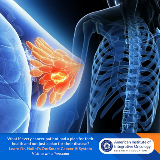
Endocrine Therapy Provides TwentyYear Benefit in ER+ Breast Cancer

Integrative and Individualized cancer care is the best cancer care and yields the best long term outcomes. Making individualized care decisions and including a health model, not just a disease model and following the OutSmart Cancer® Diet Guidelines are core principles of the OutSmart Cancer® System.

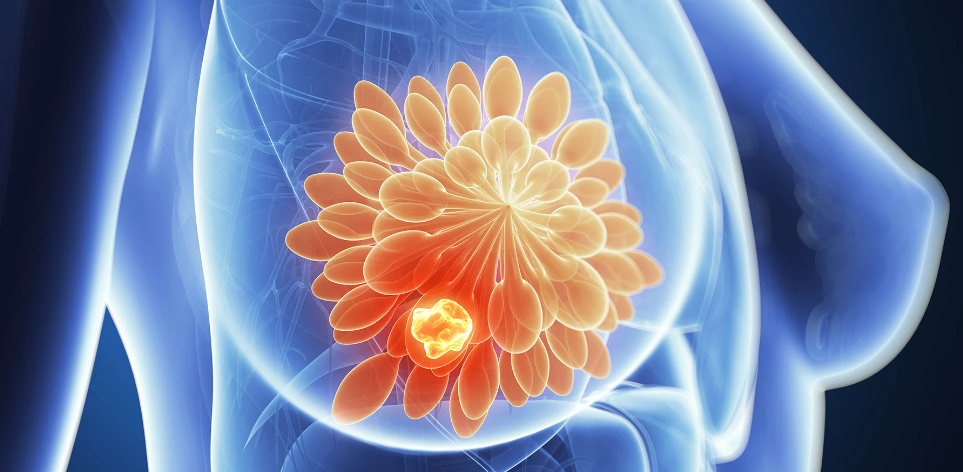
Two years of adjuvant endocrine therapy in premenopausal patients with estrogen receptor-positive (ER+) breast cancer can reduce the risk of recurrence at 20 years, according to a study published in the Journal of Clinical Oncology. (2)  This study is meaningful because many oncologists recommend five to ten years of endocrine therapy.  This study clearly demonstrates that only two years is sufficient to significantly reduce risk of breast cancer recurrence 20 years after completion of conventional oncology treatment.  This study also demonstrates that women who are candidates for and who do not receive endocrine therapy have worse outcomes. 

Furthermore this study demonstrates that women with low genomic risk should receive tamoxifen and women with high genomic risk should receive goserilin for best long term outcomes.

By including an integrative approach utilizing the health principles of the OutSmart Cancer® System, we can further manage the side effects of these treatments and support healthy function and quality of life for these women.

OUTSMART CANCER SYSTEM® Integrative Approach and Health Focussed Model

chinese-characterEspecially supportive to management of adverse menopausal effects of endocrine therapy include acupuncture therapy(3), copper free bone mineral formula and optimized Vitamin D as well as traditional Chinese Herbal Tonics

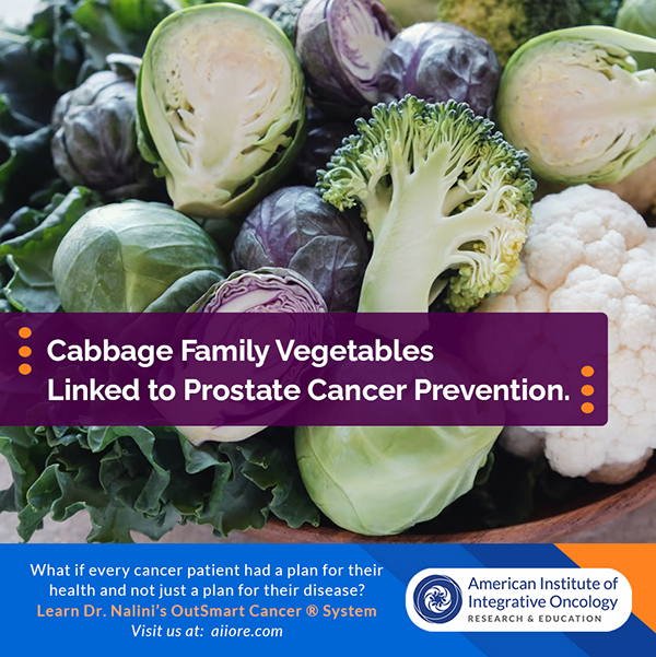
 that nourish yin and blood and support kidney qi to modulate menopausal symptoms without estrogenic effects such as Er Xian Tang (Two Immortals Formula)  and also formulas to address hormone depletion related mood dysregulation, depression and irritability such as Shu Gan Tang (Buplerum and Evodia Combination) to harmonize the liver qi and relieve stagnation of blood and emotions.  Furthermore, patients with estrogenic cancers should be given guidance on restricting estrogenic foods from their diets as part of a lifelong plan to prevent recurrence. For example,  both red meat (7) and alcohol (6) are known carcinogens linked to promotion of breast cancer and should be restricted.  Following the OutSmart Cancer® Diet Guidelines is recommended 

Monitoring for complications of endocrine therapies

Patients receiving Tamoxifen therapy  (an oral selective estrogen receptor modifier) should have semi-annual uterine ultrasound to measure endometrial thickness to assess risk of uterine hyperplasia and neoplasm as a risk of tamoxifen therapy.

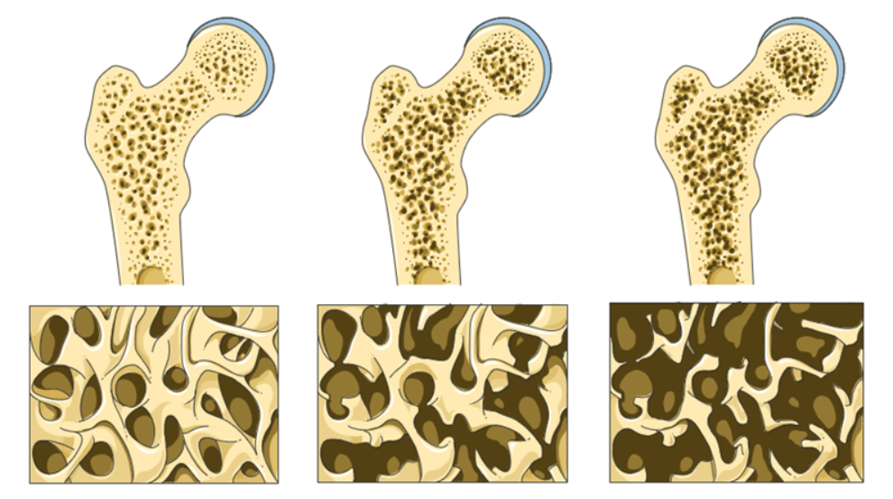
Patient receiving goserilin  (an injectabl luteinizing hormone releasing hormone antagonist administered subcutaneously either every month or every 3 months) should monitor bone density by having a baseline DEXA bone density scan at inception and at 2 years and also Urine N-Telopeptide assay to monitor rate of loss of bone minerals due to medical menopause and estrogen blockade.

Results of Study

Researchers observed significant improvements in long-term distant recurrence-free interval (DRFI) for patients who received goserelin alone, tamoxifen alone, or the combination of goserelin and tamoxifen, when compared with patients who did not receive endocrine therapy. 

However, combination goserelin and tamoxifen did not improve DRFI when compared with either agent alone.

Researchers assessed the 20-year benefit of endocrine therapy by analyzing data from the Stockholm trial (1990-1997). The analysis included 584 patients with ER+ breast cancer. The median age at baseline was 47 (range, 26-55) years, 91% of patients had progesterone receptor-positive tumors, and 88% had HER2-negative tumors.

Patients were randomly assigned to 2 years of goserelin (n=155), tamoxifen (n=135), combined goserelin and tamoxifen (n=149), or no adjuvant endocrine therapy (n=145).

In a multivariable analysis, any endocrine therapy was associated with a significant improvement in long-term DRFI, when compared with no endocrine therapy. 

There was a significant improvement in DRFI with goserelin alone (hazard ratio [HR], 0.49; 95% CI, 0.32-0.75), tamoxifen alone (HR, 0.57; 95% CI, 0.38-0.87), and goserelin plus tamoxifen (HR, 0.63; 95% CI, 0.42-0.94). 

However, there was no significant long-term benefit from the combination of goserelin plus tamoxifen, when compared with either agent alone. There was a significant interaction between goserelin and tamoxifen (P =.016). 

The researchers also assessed the long-term benefit of endocrine therapy in patients with low genomic risk (n=305) and those with high genomic risk (n=158). 

Patients with low-risk genomics had a significant improvement in DRFI with tamoxifen (HR, 0.24; 95% CI, 0.10-0.60), and patients with high-risk genomics had a significant improvement in DRFI with goserelin (HR, 0.24; 95% CI, 0.10-0.54).

Patients with high-risk genomics had significantly worse DRFI when tamoxifen was added to goserelin (HR, 3.36; 95% CI, 1.39-8.07). The interaction between goserelin and tamoxifen was significant in high-risk patients (P =.006) but not in low-risk patients (P =.080). 

“This study demonstrates long-term benefit from 2 years of adjuvant endocrine therapy in ER-positive premenopausal patients,” the researchers concluded. “Furthermore, it suggests long-lasting benefit from tamoxifen in genomic low-risk patients with long-term risk of distant recurrence, whereas genomic high-risk patients have early risk and benefit from goserelin.”

Isoflavones genistein and daidizen are phytochemicals derived from soy act  that act as MILD selective estrogen receptor modulators. These isoflavones need to be taken in very high doses on a long term basis to achieve a clinical impact and are mild in comparison to pharmaceuticals.  A recommended daily dose of genistein is 1000mg 3x/day

This study will help us support patients in making educated and informed choices and in making sure that their oncologists are practicing in accordance with the most current research and guidelines and that patients 


Using Phytochemicals

Using Phytochemicals Synergistically with Chemotherapy to Improve Efficacy and Outcomes


The role of polyphenols in overcoming drug resistance. 

In a paper published in January 2022, Maleki Dana et al, engage in a thorough review of multiple polyphenols which, when used concurrently with chemotherapy, can inhibit the development of chemo-resistance, rendering treatment more effective and for a longer duration of time.    

Most oncologists are wary of negative drug-herb, drug nutrient interactions. 


polyphenols-fruits

This review shows that we can use polyphenol phytochemicals synergistically with chemotherapy treatments to support efficacy and outcomes.


The Outsmart Cancer® System is an Integrative Cancer Care model seeking to develop highly individualized care plans that include the best therapeutic approaches and tools from multiple disciplines, combining a disease focused targeted pathology model with a whole biosystem health model to support the best outcomes for patients.


Acquired drug resistance has become a challenge that may result in treatment failure.  Multiple factors contribute to chemo-resistance in cancer cells. Acquired drug resistance occurs when cancer cells fail to respond to a previously effective treatment.  Intrinsic  chemo-resistance occurs when a pre-existing factor causes a drug to be inefficient or ineffective.


Due to the inherent heterogeneity of tumors, subpopulations of cells may develop resistance while other subpopulations remain sensitive to treatment.  

This is the rationale for using multiple agents which impact multiple signally pathways and receptors to target a wide array of heterogeneous cells.  This is also the rationale for using multiple nutriceuticals, botanicals and phytochemicals concurrently to address multiple signaling pathways and functions.

In this excellent review paper the author focuses on the multiple synergistic functions of polyphenols citing multiple studies.


Polyphenols from medicinal plants and food plants form a large part of our Materia Medica. Polyphenols include several subclasses such as catechins, flavonoids, flavones, flavonols, anthocyanins, isoflavones, curcuminoids, chalcones and phenolic acids. These natural compounds are widely found found in deeply pigmented fruits, vegetables, cocoa, seeds and green and black teas,


Not only do polyphenols inhibit multiple pathways and mechanisms of drug resistance, but also act to  confront many of the Hallmarks of Cancer, thus inhibiting and controlling the activity and viability of malignant cells.


The multiple functional roles of

  • Resveratrol
  • EGCG Epigallocatechin gallate
  • Curcumin 

are discussed and examined in depth examining multiple pathways, mechanisms of action and dosing.

Also discussed are additional well researched polyphenols, many of which are sourced from food plants as well as medicinal plants including

  • Quercetin 
  • Baicalin
  • Baicalien
  • Apigenin
  • Chrysin
  • Luteolin
  • Kaempferol

Mechanisms which lead to drug resistance in tumor cells

Malignant cells are highly adaptive and respond to toxic stressors such as chemotherapeutic drugs in the tumor micro-environment in service to their own survival.  Essential malignant cells are “smart” and they can not only co-opt normal physiologic and metabolic functions within cells to respond to the presence of toxic drug therapies.

  • Decreased Drug Uptake by tumor cells
  • Drug Efflux from tumor cells (membrane pumps)
  • Alterations in drug metabolism
  • Epigenetic Modification and Signaling
  • Inhibition of Programmed Cell death via apoptosis, autophagy and necrosis in tumor cells
  • Alterations in DNA repair 
  • Activity of Cancer Stem Cells
  • Redox Capacity of cells in response to oxidative stress
  • Alterations in Epithelial to Mesenchymal Transition, Invasion and Metastatic Progression

Not only are polyphenols capable of addressing the common Hallmarks of Cancer, but they can also inhibit the multiple ways that malignant cells seek to survive in the face of drug therapies.  


Therefore, we should strategically combine chemo-therapeutic  agents with selected polyphenols to both enhance therapeutic effect as well as inhibit the development of drug resistance. 


This leads to greater therapeutic benefit, increased duration of action as well as skillful management of adverse effects.

I encourage you to read this paper in detail for a deep and detailed review of the mechanisms of these pleomorphic multitaskers polyphenols.  The polyphenols mentioned in this paper have a history of wide use and safety.


Our role is to both eradicate and inhibit neoplastic cells as well as nurture and support the health of the patient.  This is at the heart of the OutSmart Cancer® System.


References


The Role of Polyphenols in Overcoming Cancer Drug Resistance: A Comprehensive Review 

Maleki Dana et al

Cellular and Molecular Biology Letters (2022) 27:1

https://doi.org/10.1186/s11658-021-00301-9


Hallmarks of Cancer: The Next Generation

Douglas Hanahan, Robert A. Weinberg, 2011

https://doi.org/10.1016/j.cell.2011.02.013


Revisiting the hallmarks of cancer

Fouad YA, Aanei C.. Am J Cancer Res. 2017 May 1;7(5):1016-1036. PMID: 28560055; PMCID: PMC5446472.