Home » Cancer Treatment

Category: Cancer Treatment

KNOW

Knowledge in Integrative Oncology Website

KNOW is a tool that allows access to up-to-date research on natural agents in cancer care.

KNOW: Knowledge in Integrative Oncology Website

https://www.knowintegrativeoncology.org/

Phone & Fax: 1-800-908-5175

Email info@knowoncology.org

KNOWintegrativeoncology.org is dedicated to improving the lives of people with cancer through integrative cancer care.

KNOW shares current best evidence on the use of nutrition and natural health products in oncology. Our goal is to inspire collaboration among healthcare providers, researchers, and advocacy groups to support education, safety, and clinical decision-making.

KNOW is a tool that allows access to up-to-date research on natural agents in cancer care. KNOW systematically searches and presents relevant human studies, including clinical trials, from Medline and EMBASE.

In KNOW, data is searchable by tumor type, natural therapy, conventional treatment, and side effects. You can copy references into professional communications, academic projects or presentations, education materials, curriculum, and websites. KNOW provides convenient links to the publisher for full text review or access.

Key Benefits of KNOW

  • Efficient access to current best evidence
  • Improves clinical outcomes
  • Supports development of educational resources
  • Comprehensive and cost-effective
  • Answers questions about natural therapies in cancer care

KNOW also provides Resources for Patients and Provider Network 

  • COMPETENCY AND SAFETY Articles in KNOW provide important information about safety, tolerability, preparation, dosing, and side effects not readily available to clinicians

How KNOW supports you:

✔ Improved efficiency - Enormous energy is spent to distill current literature.

✔ Stay up-to-date - The volume of research in integrative oncology is ever increasing and it's nearly impossible to stay abreast. Our team keeps the website current with summaries of published studies that the average clinician cannot easily acquire.

✔ Knowledge sharing with providers - KNOW references can be pasted into letters, handouts, presentations, and websites..

✔ Evidence-informed practice - Informed decisions require access to relevant research.

✔ Knowledge base for teaching - A central repository of information supports curriculum for integrative residencies, fellowships, and other training programs.

✔ Collaboration for research and publication projects

Membership to KNOW is subscription-based, providing access for individuals, cancer care teams, research groups, academic project groups, hospitals, and public education organizations.

For more information: https://www.knowintegrativeoncology.org/

bone cancer

Higher Risk of Bone Fracture for Cancer Survivors

Cancer stage, chemotherapy treatment, hormonal treatment, menopause status, physical activity and smoking history increase risk of bone fracture for cancer survivors.

Adult cancer survivors, specifically those who have received chemotherapy, hormonal blockade therapy and/or a diagnosis within five years, are at an increased risk for bone fractures.

bone-fracture

Recent studies published JAMA Oncology, also demonstrated decreased risk for physically active survivors and increased risk for smokers.

“These findings are important as the number of cancer survivors living in the United States is projected to rise to 26.1 million by 2040. Research like this seeks ways for cancer survivors to have a better quality of life after their diagnosis,” said Dr. Erika Rees-Punia, senior principal scientist, behavioral and epidemiology research at the American Cancer Society and lead author of the study, in a press release. “Fractures of the pelvis and vertebrae are more than just broken bones – they are serious and costly.”

Rees-Punia, et al analyzed the association between cancer stage and time of diagnosis with risk of pelvic, radial and vertebral fractures compared to adults without a history of cancer including behavior, lifestyle and type of cancer treatment. 

Among 92,431 participants included in the study, 12,943 experienced a frailty-bone fracture. Cancer survivors who were diagnosed with an advanced cancer stage within five years were at the highest risk for bone fractures compared to those without a history of cancer. Osteoporotic fractures occurred in vertebrae, pelvis and hip.

Additionally, cancer survivors who received chemotherapy had a higher rate of fracture, compared to those who did not receive chemotherapy. 

“We hope our findings will inform clinical guidance on fracture prevention, which could incorporate physical activity with exercise cancer professionals and smoking cessation programs, to improve quality of life after a cancer diagnosis,” Rees-Punia added.

Additional risks related to loss of bone density include malnutrition, persistent stress and elevated cortisol, use of steroid hormones, hyperthyroidism, estrogen and androgen hormone blockade therapies, oophorectomy, menopause, extended convalescence.

While clinicians primarily focus on risk of osteoporosis and bone fracture in women, men can also develop fracture risk and loss of bone mass. Men with low testosterone and androgens as well as men with prostate cancer being treated with androgen deprivation therapy should also be monitored for fracture risk and bone health.

Recommended Patient Guidance and Screening to reduce risk of bone fracture include

  • Bone Mineral Supplements Daily. (Copper free and including bone minerals and co factors)
  • Adequate intake of protein daily 
  • Regular weight bearing and resistance exercise
  • Active vs. Sedentary Lifestyle Support
  • Stop Smoking Support
  • Appropriate Bone Density Scans (DEXA)
  • Appropriate N-Telopeptide Crosslinks Urine Tests to assess rate of turnover of bone minerals
  • Consultation with physician to determine if anti-resorptive or hormonal                   medication may be of benefit to manage bone density and fracture risk

Selected References 

Rees-Punia E, Newton CC, Parsons HM, et al. Fracture Risk Among Older Cancer Survivors Compared With Older Adults Without a History of Cancer. JAMA Oncol. Published online November 03, 2022. doi:10.1001/jamaoncol.2022.5153

Suarez-Almazor ME, Pundole X, Cabanillas G, Lei X, Zhao H, Elting LS, Lopez-Olivo MA, Giordano SH.

Association of Bone Mineral Density Testing With Risk of Major Osteoporotic Fractures Among Older Men Receiving Androgen Deprivation Therapy to Treat Localized or Regional Prostate Cancer.

JAMA Netw Open. 2022 Apr 1;5(4):e225432. doi: 10.1001/jamanetworkopen.2022.5432.

PMID: 35363269 

Daya NR, Fretz A, Martin SS, et al. Association Between Subclinical Thyroid Dysfunction and Fracture Risk. JAMA Netw Open. 2022;5(11):e2240823. doi:10.1001/jamanetworkopen.2022.40823

Bauer DC. Clinical Use of Bone Turnover Markers. JAMA. 2019;322(6):569–570. doi:10.1001/jama.2019.9372

Prostate-Cancer-Control

Boron and Prostate Cancer Control

Aside from non-melanoma skin cancer, prostate cancer is the most common cancer among men in the United States. It is also one of the leading causes of cancer death among men of all races and Hispanic origin populations.(1)

Dietary Boron intake is inversely correlated with prostate cancer incidence. (5)

Prostate risk is  52% lower in men whose diets contain at least 1.8mg boron daily. Prostate cancer risk reduction is correlated with boron intake.  Recommended optimal daily dose of elemental boron is 3 mg/day for health maintenance.

Mechanisms of Action

Boron-containing compounds interfere with the physiology and reproduction of cancer cells through diverse mechanisms, including inhibition of serine proteases, NAD-dehydrogenases, mRNA splicing and cell division, receptor binding mimicry, and induction of apoptosis. (2) 

normal-prostate

In several studies Barranco et al (7, 8, 9, 10) demonstrate that increased boron intake is associated with lower levels of Prostate Specific Antigen (a biomarker for prostatitis and prostate cancer) as well as modulation of serum estradiol leading to increased expression of ER-beta receptors and decrease of ER-alpha receptors on tumor cells decreasing proliferation and growth signaling.  Furthermore boron supplementation increased regulation of cell cycle arrest, increased apoptosis, decreased cell adhesion, migration and metastatic progression. 

Boron decreases cancer associated inflammatory factors including hs CRP, TNFa, Interleukin-6 all of which are elevated in the tumor microenvironment.   “Elevated hs-CRP is associated with an increased risk for breast cancer, obesity and metabolic syndrome (MetS) in children, atherosclerosis, unstable angina, insulin resistance, type 2 diabetes, nonalcoholic fatty liver disease (NAFLD), metastatic prostate cancer, lung cancer, adult depression, depression in childhood and psychosis in young adult life, coronary heart disease, and stroke." (2)

In  murine study Insulin Like Growth Factor was significantly reduced in the prostate tumor cells in boron dosed animals. The IGF-1 signaling pathway promotes cancer progression and its down regulation is associated with lower risk of prostate cancer. (6)

“Increased intracellular concentration of borate activates borate transporters and leads to growth inhibition and increased apoptosis. “ (2) (3) (11)

Boron may act as a Histone De-acetylase Inhibitor (HDI).  HDI’s act as therapeutic agents for cancer due to their impact on gene expression on growth arrest signaling, cell differentiation and apoptosis in cancer cells. (2)

Boronic Acid has been shown to inhibit hypoxia inducible factor (HIF) which is a physiological stimulus for tumor induced angiogenesis.  Inhibition of HIF leads to inhibition of Vascular Endothelial Growth Factor (VEGF) and the production of capillary blood supply to tumor cells. Angiogenesis leads to exponential tumor growth and to metastatic progression of solid tumors. (11)

Plant foods rich in boron include Avocados, dried apricots, dried prunes, raisins, red kidney beans, lentils, almonds, hazelnuts, brazil nuts, pistachios, cashews and walnuts.

In summary, dietary and supplemental oral boron intake should be optimized to create a tumor microenvironment and cancer terrain that decreases risk of prostate carcinogenesis,proliferation and disease progression through angiogenesis and metastasis. 


Selected References

1. American Cancer Society Statistics https://cancerstatisticscenter.cancer.org/

2. Scorei RI, Popa R Jr. Boron-containing compounds as preventive and chemotherapeutic agents for cancer. Anticancer Agents Med Chem. 2010 May;10(4):346-51. doi: 10.2174/187152010791162289. PMID: 19912103.

3. Romulus Ion Scorei and Radu Popa, Sugar-Borate Esters –Potential Chemical Agents in Prostate Cancer Chemoprevention  DOI: 10.2174/18715206113139990124 Volume 13, Issue 6, 2013 Page: [901 - 909]

4. Kiliccioglu I, Konac E, Varol N, Gurocak S, Yucel Bilen C. Apoptotic effects of proteasome and histone deacetylase inhibitors in prostate cancer cell lines. Genet Mol Res. 2014 May 9;13(2):3721-31. doi: 10.4238/2014.May.9.17. PMID: 24854658.

5. Cui Y, Winton MI, Zhang ZF, et al. Dietary boron intake and prostate cancer risk. Oncol Rep. 2004;11(4):887-892.

6. Pizzorno, L.  Review: Nothing boring about boron. Integrative Medicine Vol. 14, No. 4 August 2015

7. Barranco WT, Hudak PF, Eckhert CD. Evaluation of ecological and in vitro

effects of boron on prostate cancer risk (United States). Cancer Causes Control. 2007;18(1):71-77.

8. Barranco WT, Eckhert CD. Boric acid inhibits human prostate cancer cell proliferation. Cancer Lett. 2004;216(1):21-29.

9. Barranco WT, Eckhert CD. Cellular changes in boric acid-treated DU-145 prostate cancer cells. Br J Cancer. 2006;94(6):884-890.

10. Barranco WT, Kim DH, Stella SL Jr, Eckhert CD. Boric acid inhibits stored Ca2+ release in DU-145 prostate cancer cells. Cell Biol Toxicol. 2009;25(4):309-320.

11. Shimizu K, Maruyama M, Yasui Y, Minegishi H, Ban HS, Nakamura H. Boron-containing phenoxyacetanilide derivatives as hypoxia-inducible factor (HIF)-1alpha inhibitors. Bioorg Med Chem Lett. 2010;20(4):1453-1456.

OutSmart-Cancer-Immuno-Therapy

Modulating Extreme Adverse Effects of Immunotherapy Treatments

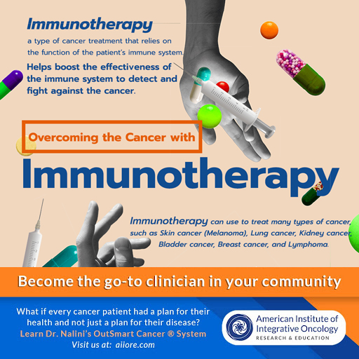
Today, more and more patients are avoiding toxic chemotherapy in favor of targeted cancer therapies.  Among the many new therapies available are a class of immunotherapy drugs that take the brakes off of the immune system and mobilize T cells against tumor cells.

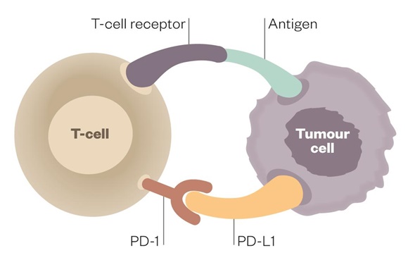
Because tumor cells have the capacity to disable T cells, this therapy addresses the huge problem of immune resistance in many cancers.  Drugs in the class of PD1 and PDL1 inhibitors were some of the first to be developed.  These drugs bind to PD1 or PDL1 receptors on the tumor surface and unleash the fury of the immune system upon the tumors by removing the inhibitory function of these ligands.

Nature has designed the immune system with both a gas pedal and a brake.  The PD1 and PDL1 inhibitors are the brakes.  Take off the brakes and the immune system is activated.

The best of outcomes with these treatments may result in complete tumor eradication, a truly miraculous outcome for some patients.  I have a patient who came to me with Stage 4 Endometrial Cancer with Lung Metastases some years ago.  After reduction of some of her tumor burden with surgery and rather brutal chemotherapy, her very forward-thinking Gynecologic Oncologist included a course of Keytruda (Pembrolizumab), which was a new immunotherapy treatment at the time. 

The historical prognosis for this patient would have been certain eventual mortality for her metastasized aggressive cancer.  However, she achieved a complete response and has been in remission and designated NED or No Evidence of Disease for many years now.   This is a patient who most likely would not be alive today without the advent of PD1 -PDL-1 inhibitor therapy.

The problem with this class of drugs is that their use is very unskillful and very unpredictable. Some patients will respond with a modicum of mild to moderate systemic autoimmune inflammation while other patients will be disabled by furious, extreme, and damaging autoimmune syndromes.  Some patients may die from extreme autoimmune activity.  I had one patient who developed myocarditis and died within a few days of receiving his first dose.  This was a prostate cancer patient whose sudden death was completely unexpected and not predicted.   

These patients require a health model and safe, effective modulation of extreme auto-immune inflammation not provided by their oncology teams. 

Some patients will have immune activation similar to a nice warm burning ember.  They get the therapeutic benefit without extreme adverse effects.  While other patients will respond with a forest fire of inflammation that must be suppressed aggressively with steroid medications for long periods of time.  The adverse effects of long-term steroid therapy then become part of the clinical picture and challenge for these patients.  In these circumstances, it IS reasonable to ask if the cure is worse than the disease itself? 

My patient developed such severe colitis (a common adverse effect) that she visited the emergency room multiple times for fluid and electrolyte replacement due to extreme persistent watery diarrhea.  Additionally, the nutritional status of this patient was also compromised and she became depleted in both calories and nutrients and developed sarcopenia.

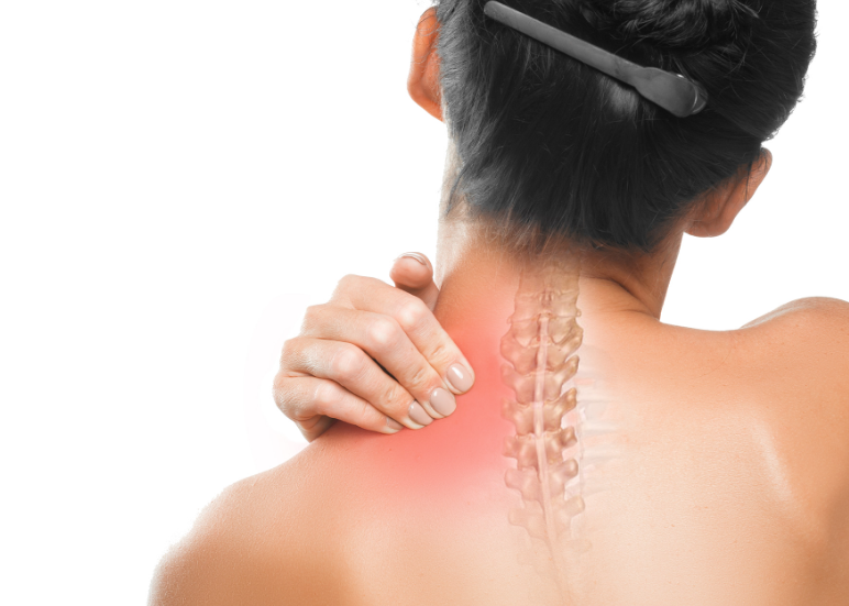
Many cancer patients receiving PD1 and PDL 1 inhibitors are left with lifelong autoimmune disease.   Most common are autoimmune arthritis, colitis, thyroiditis, dermatitis, pneumonitis, and associated loss of normal tissue and organ function.  Some patients suffer ongoing chronic inflammatory pain syndromes.

Less prevalent, but also part of the long list of adverse effects are myocarditis, pericarditis, nephritis, hepatitis, pancreatitis, neuritis, vasculitis. Essentially, any tissue or organ can be impacted with associated loss of function and sequelae.

It is my practice to screen and monitor patients receiving cancer immunotherapies for the development of autoimmune syndromes and intervene early.  If I have a patient with a history of inflammatory or autoimmune disorders I can predict that such patients are more likely to develop adverse effects. 

Additionally, high levels of inflammation not only lead to pain syndromes but are also contributors to ongoing chronic fatigue as well as agitation,  cognitive changes, sleep disruption, anxiety, and depression, and the stress of living not only as a cancer survivor but with a chronic and distressing autoimmune syndrome difficult to control and manage.  It is my firm goal that Quality of Life must be a goal in all treatment plans for cancer patients and survivors.

 If a patient has NO inflammatory adverse effects it is assumed that the patient is not going to benefit from the PD1/PDL1 inhibitor because there is no sign or symptom indicating immune activation.   I always tell patients we should celebrate if they develop a rash or diarrhea because we know the drug is working!   

In fact, it is my observation over many years of following patients who have received these therapies that when the course of immunotherapy treatment is completed those patients who continue to have low levels of inflammation continue to have the therapeutic benefit of tumor control.  This is only an empirical observation on my part.  For example, the endometrial cancer patient described above continues to have mild colitis and has remained in remission.  Before the availability of these therapies, we would expect this patient to have a recurrence and to die of her advanced stage 4 metastatic disease within a few years of her diagnosis.   Patients such as this with lung metastases historically had very poor prognoses and very high mortality rates.  Patients with powerful and manageable responses to PD1 and PDL1 inhibitors may live a long time.  While some patients do recur, some have not.  We have not had decades of time to follow these patients as these treatments are relatively new.  If nothing else, these treatments do extend the life of many patients.

How can we modulate the auto-immune adverse effects of these potentially curative immunotherapy treatments?   I have taken the approach that we employ in Functional and Naturopathic Medicine in the management of auto-immune syndromes to turn down the volume on the immune inflammation just enough to reduce extreme side effects, damage, and loss of function without losing the therapeutic benefit of these immunotherapy treatments.   

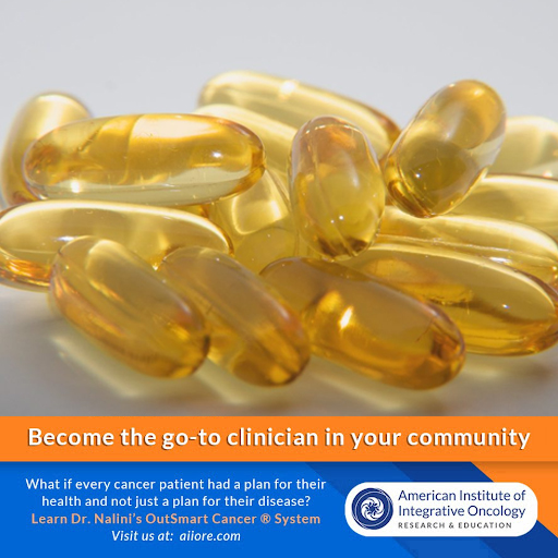
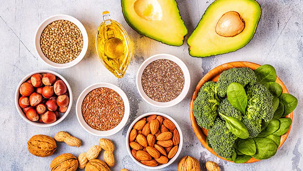
While we can rely on studies that have demonstrated that Omega 3 Fatty Acids, Vitamin D3, and Curcumin and a healthy microbiome can modulate auto-immune inflammation, there is a paucity of research on managing autoimmune syndromes related to immunotherapy adverse effects with the exception of steroids. (See selected references below.)

I share with you here my empirical clinical experience.  I have employed this approach with several hundred patients since immunotherapies have come into wider use in oncology.  Clinicians experienced in managing autoimmune syndromes will recognize the basic principles of care.

  • Anti-Inflammatory, Low Antigen Diet
  • Support for the healthy intestinal microbiome 
  • Specific Nutriceutical-Phytochemical Interventions
    • Omega 3 Fatty Acids (EPA DHA)  recommended dose 4-6 grams daily SPMs Specialized ProResolving Mediators can also be considered 1-2grams daily
    • Fat soluble Curcumin recommended dose 2-6 grams daily
    • Vitamin D3 5,000-25,000iu daily  (125mcg-625mcg). 
      • Consider a loading dose of 50,000iu (1.25mg)

I always start at the lower end of the dose range and spread the dosing out into 3-4 doses over the day.  The goal is to MODULATE but not SUPPRESS the therapeutic impact.  It is also important to be mindful of the anticoagulant/platelet aggregation inhibitory effect of such an approach and to determine which patients may NOT be a candidate for high dosing due to thrombocytopenia or anticoagulant pharmaceutical therapies.

This approach has few negative drug-nutrient interactions. I have continued these inflammation-modulating therapies continuously for many years with most patients.  Dosing is highly individualized to each patient towards the goal of supporting and promoting healthy function and quality of life.

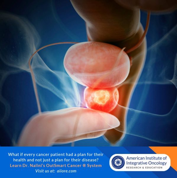
For front-line clinicians interested in supporting the health of cancer patients and cancer survivors and learning and implementing my OutSmart Cancer® System developed over 35 years in practice, I encourage you to join our training program, Foundations of Integrative Oncology, self-paced online training with clinical supervision and mentoring.  Go to aiiore.com.  

There is a huge population of patients whose lives have been touched by cancer searching for a health model and skilled and knowledgeable clinicians.

Selected References:

Vitamin D and autoimmune diseases.
Illescas-Montes R, Melguizo-Rodríguez L, et al Life Sci. 2019 Sep 15;233:116744. doi: 10.1016/j.lfs.2019.116744. Epub 2019 Aug 8. PMID: 31401314 

Vitamin D intake is associated with decreased risk of immune checkpoint inhibitor-induced colitis.
Grover S, Dougan M, et al Cancer. 2020 Aug 15;126(16):3758-3767. doi: 10.1002/cncr.32966. Epub 2020 Jun 22. PMID: 32567084

Therapeutic Potential of omega-3 Polyunsaturated Fatty Acids in Human Autoimmune Diseases.
Li X, Bi X, Wang S, Zhang Z, Li F, Zhao AZ.
Front Immunol. 2019 Sep 27;10:2241. doi: 10.3389/fimmu.2019.02241. eCollection 2019.  PMID: 31611873 

Resolvins: Emerging Players in Autoimmune and Inflammatory Diseases.
Abdolmaleki F, Kovanen PT, et al 
Clin Rev Allergy Immunol. 2020 Feb;58(1):82-91. doi: 10.1007/s12016-019-08754-9. PMID: 31267470 

Curcumin in Autoimmune and Rheumatic Diseases.
Yang M, Akbar U, Mohan C.
Nutrients. 2019 May 2;11(5):1004. doi: 10.3390/nu11051004.
PMID: 31052496 

Curcumin and autoimmune disease.
Bright JJ.
Adv Exp Med Biol. 2007;595:425-51. doi: 10.1007/978-0-387-46401-5_19. PMID: 17569223  

Curcumin as an Adjuvant to Cancer Immunotherapy.
Paul S, Sa G.
Front Oncol. 2021 Aug 16;11:675923. doi: 10.3389/fonc.2021.675923. eCollection 2021.
PMID: 34485117 

Gut Bacteria Influence Effectiveness of a Type of Immunotherapy. https://www.cancer.gov/news-events/cancer-currents-blog/2018/gut-bacteria-checkpoint-inhibitors. Feb 2018  NCI Staff

cancer-patient

VITAL TALK: Learn to Communicate Effectively with Patients Experiencing Serious Illness

“Just as no doctor is born knowing how to handle a scalpel, the same is true for how to communicate effectively with seriously ill patients and their families. We believe every clinician can become a better communicator.”

dr-tony

I met oncologist Dr. Tony Back, MD at a meditation retreat focused on working skillfully and mindfully with patients with serious, life changing illnesses as well as end of life care.  Dr. Back is affiliated with the Center to Advance Palliative Care at Fred Hutchinson Cancer Center in Seattle, Washington and has combined his years of clinical oncology experience with his recognition that patients and clinicians require more skillful and effective conversations in the midst of emotionally charged and medically complex discussions. Dr. Back has developed online and in person trainings called Vital Talk for clinicians to LEARN TO COMMUNICATE EFFECTIVELY WITH PATIENTS EXPERIENCING SERIOUS ILLNESS. His mission is “To Elevate Patient Care with better communication”.

Dr. Back and the Vital Talk mission is “that every seriously ill patient will be surrounded by clinicians who can speak about what matters most and match care to values.”

Vital Talk Trainings help clinicians to Master Tough Conversations

Whether in person or online, clinicians feel safe practicing newly learned skills through VitalTalk’s evidence-based training methodologies.

Some tips from a Vital Talk trained physician

Dr. Nalini

  • Give yourself grace
  • Lean in with emotion, heart and feeling
  • Respond with empathy.  
  • Explore and ask the patient to tell you more.  
  • Ask permission to talk about a topic (scan results, disease progression, end of life care)

[READ MORE]

EVIDENCE-BASED SKILLS Vital Talk TRAINING COURSES 

  • Elevate patient care with better communication
  • Explore best-practice communication methodologies and tools, as well as our rich community of support to better serve the needs of patients with serious illness and their families.
  • Support clinicians in learning to communicate effectively and compassionately with patients living with serious illness. 

We are living in the midst of an aging patient population in the United States. Therefore our patients are increasingly at risk of developing more serious illnesses, experiencing co-morbidities and facing many life changes of loss and limitation, frailty and mortality.   Cancer patients of all ages face immense challenges and must redefine their sense of self and examine their values and relationship to life, suffering, death and dying. These challenges may be turned into gateways to transformation.  I counsel my patients to reframe their cancer journey so that the challenges that they face become opportunities for growth, insight and wisdom and not just suffering, grief and loss.   These conversations require clinicians to develop greater communications skills and the ability to be comfortable engaging in these dialogues.

It is my experience that clinicians who are inspired to work with cancer patients are motivated by the drive towards making a difference, the intellectual challenge of the complexity of cancer and the deep meaning that arises in meeting patients at the edge where  the rawness and overwhelm of being a cancer patient can be transformed into a profound healing journey of awareness and transformation.   

I highly recommend that all clinicians continue to develop their clinical skills so that they can be fully present, capable and comfortable for participating in these most tender, human and meaningful conversations.   

I always feel it is a privilege and an honor to be allowed to accompany a patient and their family on their journey and to be able to guide them into deeper meaning and a greater sense of connection to each other and to all of life.

I encourage you to explore The Vital Talk website where you will find information on their programs and trainings but also Free Downloadable Tools and Guides to get you started

https://www.chooseyourpath.vitaltalk.org/

https://www.vitaltalk.org/resources/

Please do share your stories with us!!  

Cheating-cancer-Athena-Aktipis

How Evolution Helps Us Understand and Treat Cancer

The Cheating CellThe Cheating Cell: How Evolution Helps Us Understand and Treat Cancer - by Athena Aktipis

A fundamental and groundbreaking reassessment of how we view and manage cancer

When we think of the forces driving cancer, we don’t necessarily think of evolution. But evolution and cancer are closely linked, for the historical processes that created life also created cancer. The Cheating Cell delves into this extraordinary relationship, and shows that by understanding cancer’s evolutionary origins, researchers can come up with more effective, revolutionary treatments.


Athena Aktipis goes back billions of years to explore when unicellular forms became multicellular organisms. Within these bodies of cooperating cells, cheating ones arose, overusing resources and replicating out of control, giving rise to cancer.

Aktipis illustrates how evolution has paved the way for cancer’s ubiquity, and why it will exist as long as multicellular life does.

Even so, she argues, this doesn’t mean we should give up on treating cancer—in fact, evolutionary approaches offer new and promising options for the disease’s prevention and treatments that aim at long-term management rather than simple eradication.

Looking across species—from sponges and cacti to dogs and elephants—we are discovering new mechanisms of tumor suppression and the many ways that multicellular life-forms have evolved to keep cancer under control.

By accepting that cancer is a part of our biological past, present, and future—and that we cannot win a war against evolution—treatments can become smarter, more strategic, and more humane.

Unifying the latest research from biology, ecology, medicine, and social science, The Cheating Cell challenges us to rethink cancer’s fundamental nature and our relationship to it.

Grab Your Copy Here

fight-prostate-cancer-vegetables

Prostate Cancer Chemoprevention: I3C and DIM

Increasing serum levels of phytochemical DIM may be chemopreventive and chemoprotective for prostate cancer.

Cruciferous Cabbage Family vegetables such as (including cabbage, cauliflower, Brussels sprouts, broccoli, kale, arugula, bok choy and more ) are rich in dietary phytochemicals including Sulforaphane-Glucosinolate family molecules 13C (Indole-3-Carbinol) and it’s major bioactive therapeutic metabolite DIM  (3 3’di-indole methane).

Chopping, chewing, massaging and lightly steaming cruciferous vegetables activates the plants own catalyzing myrosinase enzyme and exposing the plant to the acid environment in the stomach leading to further metabolism resulting in bio available and bio active DIM.  DIM levels can be measured in the serum.

There are numerous studies on the benefits of dietary consumption of cruciferous vegetables.. A high intake of cruciferous vegetables is associated with reduced risk of several human cancers.  There are strong associations between high intake of broccoli and breast cancer and prostate cancer in humans.  Human cell studies show inhibition of  cell growth of several cancers including breast, prostate, pancreatic, colorectal, lung and head and neck cancers.

DIM and Prostate Cancer

DIM

DIM appears to be a potent inhibitor of human androgen hormones which may promote expression of androgen receptors on prostate cancer cells which may lead to carcinogenesis. Increasing serum levels of DIM through diet and supplementation may therefore be chemopreventive for prostate cancer.

Human and Cell Studies have shown that increased serum levels of DIM 

  • Reduces Serum Prostate Specific Antigen
  • Reduces Serum Androgen Hormones
  • Down Regulates Prostate Stem Cell Activity
  • Decreases  Nuclear Androgen Receptors
  • Induces p450 metabolic detoxification enzymes CYP1A1, CYP1A2 and CYP1B
  • Decreases oxidative stress via nrf2-KEAP pathway

The OutSmart Cancer® System is focused upon transforming the tumor microenvironment  which is a signaling environment, so that there is less physiologic support for the development and spread of cancer.   In a health model (rather than a disease model) we endeavor to transform the biosystem and  reduce pro-carcinogenic and proliferative signaling and prevent cellular, nuclear and mitochondrial damage.

brocolli

Therefore we use phytochemicals such as I3C and DIM and dietary interventions to influence carcinogenic and proliferative signaling in the tumor microenvironment.

Guidelines for Increasing Serum DIM levels

Primary Reference

*Amare DE. Anti-Cancer and Other Biological Effects of a Dietary Compound 3,3ʹ-Diindolylmethane Supplementation: A Systematic Review of Human Clinical Trials. Nutrition and Dietary Supplements. 2020;12:123-137

https://doi.org/10.2147/NDS.S261577

Additional Selected References 

  1. Anderton MJ, Manson MM, Verschoyle RD, et al. Pharmacokinetics and tissue disposition of indole-3-carbinol and its acid condensation products after oral administration to mice. Clin Cancer Res. 2004;10(15):5233–5241. doi:10.1158/1078-0432.CCR-04-0163
  2. Bjeldanes LF, Kim JY, Grose KR, et al. Aromatic hydrocarbon responsiveness-receptor agonists generated from indole-3-carbinol in vitro and in vivo: comparisons with 2, 3, 7, 8-tetrachlorodibenzo-p-dioxin. Proc Natl Acad Sci U S A. 1991;88(21):9543–9547. doi:10.1073/pnas.88.21.9543
  3. Chang Y-C, Riby J, Chang GH-F, et al. Cytostatic and antiestrogenic effects of 2-(indol-3-ylmethyl)-3, 3′-diindolylmethane, a major in vivo product of dietary indole-3-carbinol. Biochem Pharmacol. 1999;58(5):825–834. doi:10.1016/S0006-2952(99)00165-3
  4. Chen I, McDougal A, Wang F, et al. Aryl hydrocarbon receptor-mediated antiestrogenic and antitumorigenic activity of diindolylmethane. Carcinogenesis. 1998;19(9):1631–1639. doi:10.1093/carcin/19.9.1631
  5. Bradfield CA, Bjeldanes LF. High-performance liquid chromatographic analysis of anticarcinogenic indoles in Brassica oleracea. J Agric Food Chem. 1987;35(1):46–49. doi:10.1021/jf00073a010
  6. Weng J-R, Tsai C-H, Kulp SK, et al. Indole-3-carbinol as a chemopreventive and anti-cancer agent. Cancer Lett. 2008;262(2):153–163. doi:10.1016/j.canlet.2008.01.033
  7. Bradlow HL. Indole-3-carbinol as a chemoprotective agent in breast and prostate cancer. In Vivo. 2008;22(4):441–445
  8. Ahmad A, Ali S, Wang Z, et al. 3, 3′‐diindolylmethane enhances taxotere‐induced growth inhibition of breast cancer cells through downregulation of FoxM1. Int J Cancer. 2011;129(7):1781–1791. doi:10.1002/ijc.25839
  9. Ali S, Banerjee S, Ahmad A, et al. Apoptosis-inducing effect of erlotinib is potentiated by 3,3ʹ-diindolylmethane in vitro and in vivo using an orthotopic model of pancreatic cancer. Mol Cancer Ther. 2008;7(6):1708–1719. doi:10.1158/1535-7163.MCT-08-0354
  10. Banerjee S, Wang Z, Kong D, et al. 3, 3′-Diindolylmethane enhances chemosensitivity of multiple chemotherapeutic agents in pancreatic cancer. Cancer Res. 2009;69(13):5592–5600. doi:10.1158/0008-5472.CAN-09-0838
  11. Giovannucci E, Rimm EB, Liu Y, et al. A prospective study of cruciferous vegetables and prostate cancer. Cancer Epidemiol Prev Biomarkers. 2003;12(12):1403–1409.
  12. Kong D, Heath E, Chen W, et al. Loss of let-7 up-regulates EZH2 in prostate cancer consistent with the acquisition of cancer stem cell signatures that are attenuated by BR-DIM. PLoS One. 2012;7(3):e33729. doi:10.1371/journal.pone.0033729
  13. Abdelbaqi K,Lack N, Guns ET, et al. Antiandrogenic and growth inhibitory effects of ring‐substituted analogs of 3, 3′‐diindolylmethane (Ring‐DIMs) in hormone‐responsive LNCaP human prostate cancer cells. Prostate. 2011;71(13):1401–1412.
  14. Bhattacharjee S, Dashwood RH. Epigenetic Regulation of NRF2/KEAP1 by Phytochemicals. Antioxidants. 2020; 9(9):865. https://doi.org/10.3390/antiox9090865
  15. Li Y et al. Recent progress on nutraceutical research in prostate cancer. Cancer Metastasis Rev. 2014;33(2-3):629-640.
omega 3 fatty acids

Omega 3 Fatty Acids: Enhanced Control of Cancer Risk and Progression

A diet high in polyunsaturated fatty acids, especially omega 3s, have been shown to be negatively associated with cancer development

 Dietary fatty acids have been recognized as influential factors in the activation of carcinogenic events or disease progression and have been associated with a direct connection to breast cancer prevention.

PUFAs differentially inhibit mammary tumor development by inflicting modifications to the morphology of cell membranes, and influencing signaling pathways, gene expression and apoptosis.

The human body is unable to synthesize long-chain polyunsaturated fatty acids (PUFAs) Omega 3 DHA, docosahexaenoic, and EPA, Eicosapentaenoic acid and Omega 6 Arachidonic Acid at a reasonable rate and therefore, supplementation is required through dietary sources or nutritional supplements. The recommended daily nutritional dose is 2,000 mg EPA+DHA, while therapeutic dosing is 4,000-6,000 milligrams of EPA+DHA per day.

omega-3-natural

 Omega Three Fatty Acids and the Tumor Microenvironment

  1. Supports Normal Inflammation Control by lowering COX 2, LOX5, PGE2, IL1, IL6,TNFa, CRP.
    • Increased inflammation contributes to cancer development, progression and metastasis.
    • Increased inflammation is linked to cancer related pain, fatigue, depression and cognitive impairment.
    • Increased inflammation is linked to cancer related hypercoagulation and risk of thromboembolism
    • Supporting Normal Inflammation control has a wide impact on the behavior of tumor cells and on safety and quality of life for cancer patients and survivors.
  2. Promotes Expression of M1 Type Tumor Associated Macrophages (TAMs).
    • Type M1 TAMs promote tumor regression, inflammation control and immune activation by promoting tumor infiltration by antigen presenting dendritic cells and cytotoxic T cells.
  3. Inhibits VEGF (Vascular Endothelial Growth Factor) and Promotes Normal Control of Angiogenesis .
    • VEGF promotes the development of new blood vessels to the tumor cells. Inhibition of VEGF and the development of capillaries inhibits tumor growth and profession as well as metastasis.
       
  4. Down regulates tumor promoter Protein Kinase C isoenzymes,
    • A group of enzymes that link multiple cellular processes responsible for regulation of tumorigenesis, cell cycle progression and metastasis.
  5. Inhibits Collagenase,
    • A proteolytic enzyme that breaks down the ECM (Extracellular Matrix) and allows invasion of tumor cells into tissues and blood vessels, leading to progression, invasion and metastasis.
  6. Promotes Normal Apoptosis signaling.
    • Cancer cells lose the ability to initiate apoptosis, the normal process in which a cell recognizes itself as aberrant and self destructs. The inhibition of normal apoptotic signaling in malignant cells is a hallmark  of the tumor microenvironment permissive of uncontrolled growth, persistence and immortality due to loss of normal regulation.
  7. Lowers Bcl2 and Ras oncogenes.
    • These genes inhibit normal apoptosis and promote tumor growth and progression.
  8. Acts as a Chemo-sensitizer
    • Working synergistically to enhance therapeutic effect of chemotherapy drugs. DHA has a potential to specifically chemo-sensitize tumors.
    • Tumour cells can be made more sensitive to chemotherapy than non-tumor cell when membrane lipids are enriched with DHA
    • Incorporating DHA during treatment reduces adverse effects of chemotherapy.
    • DHA can improve the outcome of chemotherapy when highly incorporated into cell membranes.
  9. Acts as a Radio-sensitizer.
    • By promoting normal membrane structure and function and by influencing the tumor microenvironment DHA acts synergistically to potentiate therapeutic effects of radiotherapy on tumor cells.
  10. Promotes Healthy 16-OH Estrogen metabolism.
    • Estrogen can be metabolized through multiple pathways. The promotion of 16-Hydroxylation of estrogen produces estrogen metabolites that are not pro-carcinogenic. Omega 3 Fatty Acids promote healthy estrogen metabolism.
  11. Inhibits Platelet Aggregation and Thrombin Formation.
    • Abnormal hyper-coagulation, increased platelet aggregation and thrombus formation are hallmarks of the tumor microenvironment. Control of platelet aggregation and thrombus formation reduces the risk of life threatening and adverse  thrombotic events.  40% of all cancer patients are at risk for the formation of thromboembolisms.  Omega 3 Fatty Acids reduce this risk.
  12. Promotes Normal Cell Membrane Functions and Receptor Binding
    • A healthy flexible cell membrane built of omega 3 fatty acids promotes an enhancement of all membrane functions, normalizing and optimizing normal and therapeutic physiology.
  13. Increases expression of Tumor Suppressor Gene PTEN.
    • Increased expression of tumor suppressor genes leads to enhanced control over carcinogenesis,  tumorigenesis and metastatic progression.
  14. Inhibits Multi Drug Resistance.
    • Tumor cells can quickly become resistant to therapeutic anti-neoplastic agents thus decreasing and shortening the efficacy of treatments.
  15. Inhibits cachexia preserves muscle mass and bone mass (inhibits proteolysis inducing factor)
    • Loss of bone mass (osteopenia) and loss of muscle mass (sarcopenia) are risk factors of aging and of the cancer physiology.  Maintaining bone mass and muscle mass are crucial to robust healthy function and quality of life.
  16. Supports normal mood regulation.
    • Depression and anxiety are common in cancer patients. Support of balanced mood allows cancer patients deep and restful sleep, improved quality of life and increased coping capacity and resilience in the face of stress.

Cautions and Contraindications

  • Patient on anticoagulant medications
  • Patients with thrombocytopenia and known hypo-coagultion clotting disorders
  • Pre and Post Surgical patients (72 hours)
  • Patients with seafood allergies


How to Measure Omega 3 Fatty Acid Status

Serum or Plasma Omega 3 Fatty Acid ratios. LABCORP Omega 3-6 Fatty Acids, Quest Diagnostics Omegacheck, Boston HeartLab Fatty Acid Balance, Cleveland HeartLab Omegacheck, Genova Diagnostics Essential and Metabolic Fatty Acids, Great Plains Comprehensive Fatty Acids, OmegaQuant Omega3 Index.

Selected References

 Azrad M, Turgeon C, Demark-Wahnefried W. Current evidence linking polyunsaturated Fatty acids with cancer risk and progressionFront Oncol. (2013) 3:224.

 Bartsch H, Nair J, Owen RW. Dietary polyunsaturated fatty acids and cancers of the breast and colorectum: emerging evidence for their role as risk modifiers. Carcinogenesis. (1999) 20:2209–18.

 Bournoux, P. Et al. Improving outcome of chemotherapy of metastatic breast cancer by DHA: Phase II Trial, Br.J Cancer 2009 Dec 15:101(12):1978-85

 Shweta Tiwary   Altered Lipid Tumor Environment and Its Potential Effects on NKT Cell Function and Tumor Immunity.  Front Immunol.10.3389/fimmu.2019.02187

 Zanoaga O, Jurj A, Raduly L, Cojocneanu-Petric R, Fuentes-Mattei E, Wu O, et al. Implications of dietary omega-3 and omega-6 polyunsaturated fatty acids in breast cancer.  Exp Ther Med. (2018) 15:1167–76. 10.3892/etm.2017.5515

MRI

Innovative MRI without Toxic Gadolinium Contrast Media

PreNuvo: Innovative MRI Technology

High-resolution radiology is used in oncology for both diagnoses as well as screening and monitoring. Because cancer cell physiology is metabolically different from healthy cells, contrast media are preferentially taken up by cancer cells allowing for more precise imaging.

Magnetic Resonance Imaging (MRI)  was invented in the 1980s.  It is a magnetic technology and does not expose patients to ionizing radiation, but to magnetic fields.  Hence it is considered safer than exposure to the damaging, oncogenic ionizing radiation found in X-Rays and PET and CT scans

Say No to Gadolinium
The most commonly used contrast medium used with MRI imaging to enhance resolution is Gadolinium. Gadolinium is a  magnetic metal that is engineered into a nanoparticle solution and injected into the vein.

Gadolinium can damage both nephrons and neurons and is not completely excreted leading to toxic load over time.  Because cancer patients may have multiple scans per year over many years, exposure to Gadolinium can become damaging and increasingly toxic. Gadolinium also acts as a calcium channel blocker and even at low concentrations can interfere with the contraction of smooth, skeletal, and cardiac muscle, nerve impulses, and blood coagulation. 

MRI-Man

Furthermore, there is a syndrome called Gadolinium deposition disease that is a gadolinium storage condition for which the long-term effects are not well understood.  A common adverse effect of Gadolinium exposure and retention is renal fibrosis.

Patients should discuss the risks of ALL contrast media with the radiology team to evaluate risks and benefits and be fully informed before proceeding with any scan.  Because all contrast media has toxicity, the opportunity to have a high-resolution scan without the use of Gadolinium contrast media is a very important innovation.

Patients with compromised cardiovascular, renal and neurologic function should use cautions before authorizing the performance of any scan without a full understanding of toxicity, risks, and benefits of contrast media

What if A High-Resolution MRI was possible without toxic contrast media?

PreNuvo has developed a High-Resolution MRI Scan that does not require the use of Gadolinium contrast media.

As with all technologies, MRI technology is advancing. Prenuvo is a company on the forefront of MRI Innovation bringing new safe and effective techniques not requiring contrast medium for high-resolution whole-body imaging, making accurate diagnosis possible.

Prenuvo uses innovative new hardware, software, and sequencing to create more detailed comprehensive images along with the use of Artificial Intelligence to enhance accurate analysis in less time.  Prenuvo claims a 0.7% false-positive rate due to the higher resolution, often decreasing the need for additional follow-up imaging, biopsies, or surgeries.  All without the use of Gadolinium or any other contrast media.

A more comfortable patient-centric experience:
A whole-body scan at Prenuvo typically takes 60 minutes compared to 5 hours for a conventional MRI.  The scanner itself is wider and more open and less claustrophobic with fresh air vents.  The machine is much quieter as opposed to the loud clanking of the conventional MRI.  The design also allows the patient’s head to remain outside of the scanner for most of the scan.  Innovative design and technology also solve many of the issues that lead to stress and anxiety for the patient that is common with MRI scans.  Additionally, a headset and a music menu are provided to support relaxation.

For a faster, safer, less toxic, and highly accurate MRI without contrast, I recommend that you explore the use of prenuvo.com for your patients.

(NB: I have no financial relationship whatsoever with  Prenuvo.)

Selected References:

copper-drive

How does Copper Drive Cancer Progression?

Key Takeaways:

This initiation of Angiogenesis is one of the steps of disease progression and acceleration, Creating an environment where this is contained is very powerful.

  • Controlling Copper and Angiogenesis is a mechanism
  • No one ever dies of a primary tumor for the most part, it is metastatic disease that kills the cancer patients
  • The more we can contain tumor progression, travel and desize, patients can live with metastatic disease for quite a long time, if we can control it
  • There's no kill rate to the tumor cell when you inhibit Angiogenesis, by any means whether it's a drug or copper culation
  • If we lower copper in a patient whose had a good reduction of their tumor burden, by any means, surgery, chemotherapy and immunotherapy... And then we track their copper, keeping copper in the lowest quartile of normal, usually their disease does not progress.
  • You reduce Inflammation when you reduce copper

cancer-patient-cancer-doctor