Home » Cancer Risk

Tag: Cancer Risk

breast-cancer

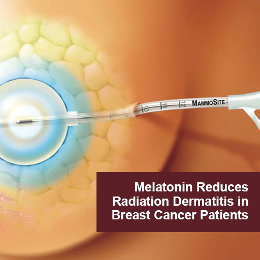
Melatonin Reduces Radiation Dermatitis in Breast Cancer Patients

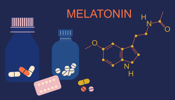
Radiation induced dermatitis is a common adverse effect and complication of radiotherapy.  Topical melatonin has proven to be a safe and effective agent in the prevention and treatment of radio-dermatitis in breast cancer patients.   Melatonin protects the skin by acting both as a topical anti-inflammatory and as an antioxidant. (5)

In a randomized placebo controlled trial women undergoing radiotherapy applied topical melatonin gel twice daily and for 2 weeks after completing radiotherapy.  The control group received a placebo gel. In the melatonin group  the occurrence of Grade1/2 radiation dermatitis was 59% vs 90% of the placebo group. “Patients treated with melatonin-containing emulsion experienced significantly reduced radiation dermatitis compared to patients receiving placebo.” (1). 

Topical Melatonin

  • Reduces inflammation and oxidative stress at the site of  irradiation
  • Delays onset of radio-dermatitis
  • Decreases the intensity of radiodermatitis and skin damage
  • Promotes resolution of dermatitis 
  • Promotes healing of skin
  • Improves Quality of Life 

Stress, nervous strain.Sleepless problem.Medicine diffuser.Flat vector

In two double blind randomized placebo controlled trials by the same research team, breast cancer patients applied a low dose melatonin cream twice daily during their course of radiotherapy.  The control group received a placebo cream.  During the first two weeks of therapy there was no difference between the 2 groups. Although there was no statistical difference, the researchers state that melatonin “demonstrated a protective effect”.   They posited that another study with a higher dose of melatonin is warranted. (3)  Researchers reported that melatonin decreased  breast symptoms of radiation dermatitis and improved quality of life for patients in the melatonin group (4)

In a case report a breast cancer patient applied melatonin cream daily and for 3 weeks after completing. radiotherapy treatment.  Radiation dermatitis did not appear at the radiated site until one week after final treatment (a delayed response) and resolved within 3 weeks of continued use of topical melatonin. (2)

There have been no reports of adverse effects with the use of  topical melatonin.

Apply topical melatonin to irradiated skin twice daily during radiotherapy and continuing for 2-3 weeks after completion of course of treatment until the skin is healed.

Melatonin gel or cream can be made by a compounding pharmacist.  Melatonin is also commercially available as a topical cream, as a liposomal solution and as a topical spray. 

References

1 Melatonin for Prevention of Breast Radiation Dermatitis: A Phase II, Prospective, Double-Blind Randomized Trial.

Ben-David MA, Elkayam R, Gelernter I, Pfeffer RM.

Isr Med Assoc J. 2016 Mar-Apr;18(3-4):188-92.

PMID: 27228641 Free article. Clinical Trial. 

2 Compounded Melatonin Cream for the Prevention and Treatment of Radiation Dermatitis: A Case Report.

Garcia-Segura LC, Garcia-Segura JC, Delgado DC, Romero MN, Salgado EC, Llorens LP.

Int J Pharm Compd. 2022 Jan-Feb;26(1):6-8.

PMID: 35081038 

3 Effect of melatonin cream on acute radiation dermatitis in patients with primary breast cancer: A double-blind, randomized, placebo-controlled trial.

Zetner D, Kamby C, Christophersen C, Gülen S, Paulsen CB, Piga E, Hoffmeyer B, Mahmood F, Rosenberg J.

J Pineal Res. 2023 Apr 13:e12873. doi: 10.1111/jpi.12873. Online ahead of print.

PMID: 37055944 


blood-test

What Common Blood Tests Can Detect Early Signs of Cancer

While there is no simple blood test for predicting who will get cancer, there is a lot of information to be gleaned from basic blood work that, taken together, reveals much about an individual’s predispositions for many forms of cancer. By monitoring selected biomarkers routinely measured in primary care, you can learn a lot about physiological patterns that promote carcinogenesis, proliferation, progression, and recurrence long before tumor markers emerge or there are radiological or pathological findings indicating cancer.

The art of assessment lies in part in recognizing the patterns. By learning how to read the multiple biochemical signals that emerge from a pro-carcinogenic “tumor microenvironment,” you can begin to practice real prevention, and give your patients the opportunity for significant improvements in both health-span and lifespan.

The tests included in this article here are ones you are routinely ordering in the integrative and functional medicine setting. While they are not to be misconstrued as diagnostic tests for cancer, they can indicate that a patient is at increased risk, and that further assessment and action is required to identify potential malignancy.

In people who’ve had cancer, these common tests are often prognostic for disease progression and recurrence.

It is vital that primary care practitioners do a better job of recognizing the early signs of recurrence among cancer survivors. According to the American Cancer Society’s 2016-2017 Survivorship Facts and Figures, the population of cancer survivors will increase to 20.3 million by January 1, 2026.

After conventional oncology treatment is finished, these patients typically return to their primary care physicians. They are highly motivated, ripe for change, and in search of clinicians who can support their efforts to restore health and prevent recurrence.

The tests described below will help you fill that role.

Complete Blood Count
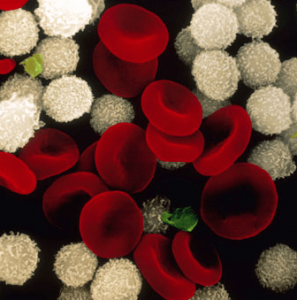
One of the most common biomarkers of overall health is the Complete Blood Cell panel, which can be used to monitor hematologic abnormalities caused by solid tumors, hematologic malignancies, as well as the side-effects of the therapies used to treat them.

The following findings are not definitive diagnostic signals, but taken together, they suggest that someone is at greatly increased risk:

  • Elevated White Blood Cells > 11.0 109/L
  • Elevated Platelets > 350 109/L
  • Low Hemoglobin <10.0 g/dL
  • High Neutrophil to Lymphocyte Ratio (NLR)

The latter finding—a high NLR—is especially important.

Neutrophils promote cancer progression, proliferation, and metastasis by increasing vascular endothelial growth factor (VEGF), Hepatocyte growth factors, inflammatory cytokines IL-6, IL-8, matrix metalloproteinases (MMP), and elastase. Neutrophils and macrophages secrete tumor growth promoting factors and contribute to a proliferative tumor microenvironment.

Therefore a high neutrophil count is suggestive of a neoplastic process somewhere in the body.

According to a 2014 metanalysis of 57 studies, an NLR greater than 4.0 was associated with a hazard ratio for overall survival (OS) of 1.81 (95% CI = 1.67 to 1.97; P < .001), an effect observed in all disease subgroups, sites, and stages and that predicts increased risk of mortality (Templeton AJ, et al. JNCI. 2014:106(6).)

Simply put, an NLR over 4 predicts tumor progression and poor overall survival. This is a readily available and inexpensive biomarker with a lot of prognostic value.

Hyperglycemia

A fasting glucose in the range of 100-126 mg/dl is suggestive of cancer risk.

Glucose may have a direct role in cancer development. Tumor cells have increased numbers of receptors for insulin, insulin-like growth factor, and GLUT4. Thus, they transport more glucose into themselves, and this promotes growth and proliferation. It is the main reason for using a low-glycemic, modified ketogenic diet in patients with cancer.

Proliferating tumor cells have up- regulated glucose transporters. Elevated serum glucose is linked to increased risk and progression of many solid cancers, including breast cancer (Haseen SD, et al. Asian Pac J Cancer Prev, 2015:  16, 675-8).

High glucose levels also result in a state of chronic inflammation, which leads to an increase of cytokines, such as interleukin 6 (IL-6), tissue necrosis factor alpha (TNF-α) and vascular endothelial growth factor (VEGF). All of these promote cancer progression, proliferation, and metastasis (Crawley DJ, et al. BMC Cancer, 14(1), 985).

Given the high prevalence of diabetes, metabolic syndrome, and insulin resistance in the US, this is an important indicator to watch.

Serum glucose is a modifiable risk factor. Diet and lifestyle changes that reduce and regulate glucose will also help to reduce risk and progression of cancer.

High Insulin & Low SHBG 

Prolonged hyperinsulinemia leads to reduced hepatic production of sex hormone binding globulin (SHBG). This, in turn, increases risk of steroid hormone driven cancers. Low SHBG results in increased amounts of unbound estrogens and androgens that drive carcinogenesis in breast, endometrial, prostate lung, colorectal and pancreatic tissues.

Free unbound estrogen also exerts immunosuppressive effects in the tumor microenvironment, and has a profound impact on anti-tumor immunity and tumor-promoting inflammation that is completely independent from its direct activity on tumor cells (Svoronos N, et al. Cancer Discovery, 2017: 7(1), 72-85).

Low Serum Albumin
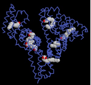
Serum albumin levels have prognostic significance in cancer, and can be used to better define baseline risk in cancer patients. It is generally useful in assessing the nutritional status, disease severity, disease progression, and prognosis.

In a multivariate analysis of 29 studies, Gupta and Lis found, “higher serum albumin levels to be associated with better survival.” (Gupta D, Lis CG. Nutrition Journal, 2010: 9(1), 69).

In the early stages of cancer, there is slight or no hypoalbuminemia. But as the disease progresses, malnutrition and inflammation suppress albumin synthesis, and albumin levels drop significantly.

Albumin levels under 3.5 g/dL are often seen in patients with sarcopenia and cachexia. Malnutrition is a predictor of reduced survival. It is also associated with deteriorating quality of life, decreased response to treatment, increased risk of chemotherapy-induced toxicity, and a reduction in cancer survival.

On the high side, albumin concentrations above 37.5 g/L are predictive of both chemotoxicity and of survival (Srdic D, et al. Supportive Care in Cancer, 2016: 24(11), 4495-4502).

It is also important to look at the Albumin-to-Globulin Ratio.

A ratio of less than 1.66 is a risk factor for cancer incidence and mortality, both short- and long term, in generally healthy screened adults. In people who’ve already developed some form of cancer, a low albumin-to-globulin ratio predicts low overall survival (Suh B, et al. Ann Ocol (2014): 25(11), 2260-2266).

Elevated Ferritin
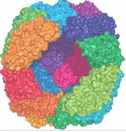
Ferritin, a strong negative survival predictor, has been associated with the pathological processes of inflammation and infection. High ferritin is suggestive of inflammation, immunosuppression, tumor angiogenesis, and proliferation.

Elevated serum ferritin—indicated by levels over 200 ng/ml in men, and over 150 ng/ml in women–have been seen in people with breast cancer, pancreatic cancer, non-small cell lung cancer, hepatocellular carcinoma, leukemia, colorectal cancer and lymphoma.

High ferritin levels are significantly associated with reduced survival time and increased mortality in cancer patients (Lee S, et al. J Cancer, 2016: 7(8), 957-964)

25-OH Vitamin D Deficiency

Vitamin D has a multi-functional impact on the tumor microenvironment. Increased levels of Vitamin D are associated with reduced occurrence and reduced mortality of different types of cancer, including skin, prostate, breast, colon, ovary, kidney, and bladder.

Vitamin D is involved in a very wide range of physiological processes relevant to cancer development, including: Regulation of Gene Transcription; Growth Arrest; Apoptosis; Cellular Differentiation; DNA Repair; Antioxidant Protection; Immune Modulation; Regulation of Pro-Inflammatory Cytokines; and Control of Angiogenesis & Metastasis.

Low or suboptimal levels of 25-OH Vitamin D are associated not only with increased risk of various forms of cancer, but also with poor prognosis, and more aggressive disease (McDonnell SL, et al. PloS One, 2016: 11(4), e0152441).

This is particularly true in breast cancer. In one study, vitamin D-deficient women with breast cancer typically had more aggressive molecular phenotypes and worse prognostic indicators than those with adequate vitamin D (Williams JD, et al. Endocrinology, 2016: 157(4), 1341-1347).

The Vitamin D Council suggests repletion to 40 to 80 ng/mL, with a target of 50 ng/ml, for optimal health on multiple fronts, including colorectal cancer prevention (Bischoff-Ferrari HA, et al. Am J Clin Nutr, 2006: 84(1), 18-28).

Supplementation to reach mean serum concentrations of 72 nmol/L showed a beneficial effect  against cancer development (Lappe JM, et al. Am J Clin Nutr. 2007: 85(6), 1586-1591).

When assessing patients in the context of cancer risk, the following guidelines are useful:

25 –hydroxy- Vitamin D (ng/ml)

Deficient                                                        < 50

Optimal                                                           50-70

Optimal for Cancer & CVD                70-99

Excess                                                            >100

Elevated Lactic Acid Dehydrogenase

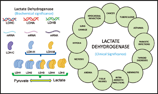
Lactate dehydrogenase (LDH) is an enzyme that catalyzes the reduction of pyruvate to lactate.

Aberrant metabolism and inefficient fuel production is a characteristic of tumor cells, which are dominated by aerobic glycolysis, increased lactate production, and a higher uptake of glucose (the Warburg effect).

Elevated LDH may be a marker of these aberrant metabolic processes in cancer cells.

The normal range for LDH is thought to be 100-333 u/L, with levels greater than 245 u/L considered to be in the upper quartile of normal. Above that 245 u/L mark, it is suggestive of early carcinogenesis, tumor cell proliferation, tumor progression, and poor prognosis.

It is often highly elevated in aggressive forms of cancer and hematological malignancies including: melanoma, lymphoma, acute leukemia, seminoma germ cell, pancreatic, gastric, lung, renal cell, nasopharyngeal, esophageal, cervical, and prostate cancers (Wulaningsih W, et al. Br J Cancer. 2015:113(9). Zhang J, et al. Sci Rep. 2015:5, 9800).

Elevated C-Reactive Protein
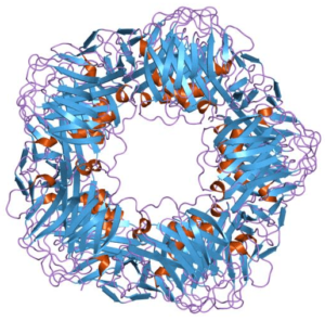
C-Reactive Protein (CRP) is a well-established inflammatory marker. It is also a biomarker of cancer survival.

CRP is elevated in patients with solid tumors, and high levels predict poor prognosis, blunted treatment response, as well as tumor recurrence.

As part of the systemic inflammatory response to a tumor, the body releases pro-inflammatory cytokines and growth factors. Interleukin-6, produced by the tumor or surrounding cells, stimulates liver production of acute-phase reaction proteins that increase C-reactive protein (CRP) and fibrinogen.

Elevated CRP correlates with disease stage and increased cancer mortality (Shrotriya S, et al. PloS One. 2015: 10(12), e0143080). Individuals with a high baseline CRP (>3 mg/L) have an 80% greater risk of early death compared with those with low CRP levels (<1 mg/L).  

Patients with invasive breast cancer and CRP levels>3 mg/L at diagnosis have a 1.7 fold increased risk of death compared to those with CRP levels<1 mg/L at diagnosis (Allin KH, et al. Breast Cancer Res. 2011: 13(3), R55).

Converging Signals

No one of the aforementioned test parameters is, in and of itself, an indicator that someone has cancer. But by looking at standard blood test results in a new way, you can start to recognize the patterns of high risk and active cancer physiology. This is crucial to early identification and early intervention.

Clinicians who are aware of the converging signs can meaningfully shift the microenvironment from one that promotes cancer to one that is not supportive of carcinogenesis, proliferation, or progression. In the same way, we can provide meaningful support for the rising tide of underserved cancer survivors and at-risk patients in need of not only a disease plan, but also a health plan.

Prostate-Cancer-Control

Boron and Prostate Cancer Control

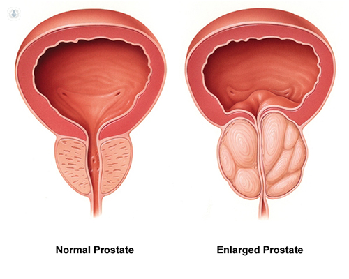
Aside from non-melanoma skin cancer, prostate cancer is the most common cancer among men in the United States. It is also one of the leading causes of cancer death among men of all races and Hispanic origin populations.(1)

Dietary Boron intake is inversely correlated with prostate cancer incidence. (5)

Prostate risk is  52% lower in men whose diets contain at least 1.8mg boron daily. Prostate cancer risk reduction is correlated with boron intake.  Recommended optimal daily dose of elemental boron is 3 mg/day for health maintenance.

Mechanisms of Action

Boron-containing compounds interfere with the physiology and reproduction of cancer cells through diverse mechanisms, including inhibition of serine proteases, NAD-dehydrogenases, mRNA splicing and cell division, receptor binding mimicry, and induction of apoptosis. (2) 

normal-prostate

In several studies Barranco et al (7, 8, 9, 10) demonstrate that increased boron intake is associated with lower levels of Prostate Specific Antigen (a biomarker for prostatitis and prostate cancer) as well as modulation of serum estradiol leading to increased expression of ER-beta receptors and decrease of ER-alpha receptors on tumor cells decreasing proliferation and growth signaling.  Furthermore boron supplementation increased regulation of cell cycle arrest, increased apoptosis, decreased cell adhesion, migration and metastatic progression. 

Boron decreases cancer associated inflammatory factors including hs CRP, TNFa, Interleukin-6 all of which are elevated in the tumor microenvironment.   “Elevated hs-CRP is associated with an increased risk for breast cancer, obesity and metabolic syndrome (MetS) in children, atherosclerosis, unstable angina, insulin resistance, type 2 diabetes, nonalcoholic fatty liver disease (NAFLD), metastatic prostate cancer, lung cancer, adult depression, depression in childhood and psychosis in young adult life, coronary heart disease, and stroke." (2)

In  murine study Insulin Like Growth Factor was significantly reduced in the prostate tumor cells in boron dosed animals. The IGF-1 signaling pathway promotes cancer progression and its down regulation is associated with lower risk of prostate cancer. (6)

“Increased intracellular concentration of borate activates borate transporters and leads to growth inhibition and increased apoptosis. “ (2) (3) (11)

Boron may act as a Histone De-acetylase Inhibitor (HDI).  HDI’s act as therapeutic agents for cancer due to their impact on gene expression on growth arrest signaling, cell differentiation and apoptosis in cancer cells. (2)

Boronic Acid has been shown to inhibit hypoxia inducible factor (HIF) which is a physiological stimulus for tumor induced angiogenesis.  Inhibition of HIF leads to inhibition of Vascular Endothelial Growth Factor (VEGF) and the production of capillary blood supply to tumor cells. Angiogenesis leads to exponential tumor growth and to metastatic progression of solid tumors. (11)

Plant foods rich in boron include Avocados, dried apricots, dried prunes, raisins, red kidney beans, lentils, almonds, hazelnuts, brazil nuts, pistachios, cashews and walnuts.

In summary, dietary and supplemental oral boron intake should be optimized to create a tumor microenvironment and cancer terrain that decreases risk of prostate carcinogenesis,proliferation and disease progression through angiogenesis and metastasis. 


Selected References

1. American Cancer Society Statistics https://cancerstatisticscenter.cancer.org/

2. Scorei RI, Popa R Jr. Boron-containing compounds as preventive and chemotherapeutic agents for cancer. Anticancer Agents Med Chem. 2010 May;10(4):346-51. doi: 10.2174/187152010791162289. PMID: 19912103.

3. Romulus Ion Scorei and Radu Popa, Sugar-Borate Esters –Potential Chemical Agents in Prostate Cancer Chemoprevention  DOI: 10.2174/18715206113139990124 Volume 13, Issue 6, 2013 Page: [901 - 909]

4. Kiliccioglu I, Konac E, Varol N, Gurocak S, Yucel Bilen C. Apoptotic effects of proteasome and histone deacetylase inhibitors in prostate cancer cell lines. Genet Mol Res. 2014 May 9;13(2):3721-31. doi: 10.4238/2014.May.9.17. PMID: 24854658.

5. Cui Y, Winton MI, Zhang ZF, et al. Dietary boron intake and prostate cancer risk. Oncol Rep. 2004;11(4):887-892.

6. Pizzorno, L.  Review: Nothing boring about boron. Integrative Medicine Vol. 14, No. 4 August 2015

7. Barranco WT, Hudak PF, Eckhert CD. Evaluation of ecological and in vitro

effects of boron on prostate cancer risk (United States). Cancer Causes Control. 2007;18(1):71-77.

8. Barranco WT, Eckhert CD. Boric acid inhibits human prostate cancer cell proliferation. Cancer Lett. 2004;216(1):21-29.

9. Barranco WT, Eckhert CD. Cellular changes in boric acid-treated DU-145 prostate cancer cells. Br J Cancer. 2006;94(6):884-890.

10. Barranco WT, Kim DH, Stella SL Jr, Eckhert CD. Boric acid inhibits stored Ca2+ release in DU-145 prostate cancer cells. Cell Biol Toxicol. 2009;25(4):309-320.

11. Shimizu K, Maruyama M, Yasui Y, Minegishi H, Ban HS, Nakamura H. Boron-containing phenoxyacetanilide derivatives as hypoxia-inducible factor (HIF)-1alpha inhibitors. Bioorg Med Chem Lett. 2010;20(4):1453-1456.

OutSmart-Cancer-Immuno-Therapy

Modulating Extreme Adverse Effects of Immunotherapy Treatments

Today, more and more patients are avoiding toxic chemotherapy in favor of targeted cancer therapies.  Among the many new therapies available are a class of immunotherapy drugs that take the brakes off of the immune system and mobilize T cells against tumor cells.

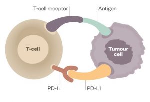
Because tumor cells have the capacity to disable T cells, this therapy addresses the huge problem of immune resistance in many cancers.  Drugs in the class of PD1 and PDL1 inhibitors were some of the first to be developed.  These drugs bind to PD1 or PDL1 receptors on the tumor surface and unleash the fury of the immune system upon the tumors by removing the inhibitory function of these ligands.

Nature has designed the immune system with both a gas pedal and a brake.  The PD1 and PDL1 inhibitors are the brakes.  Take off the brakes and the immune system is activated.

The best of outcomes with these treatments may result in complete tumor eradication, a truly miraculous outcome for some patients.  I have a patient who came to me with Stage 4 Endometrial Cancer with Lung Metastases some years ago.  After reduction of some of her tumor burden with surgery and rather brutal chemotherapy, her very forward-thinking Gynecologic Oncologist included a course of Keytruda (Pembrolizumab), which was a new immunotherapy treatment at the time. 

The historical prognosis for this patient would have been certain eventual mortality for her metastasized aggressive cancer.  However, she achieved a complete response and has been in remission and designated NED or No Evidence of Disease for many years now.   This is a patient who most likely would not be alive today without the advent of PD1 -PDL-1 inhibitor therapy.

The problem with this class of drugs is that their use is very unskillful and very unpredictable. Some patients will respond with a modicum of mild to moderate systemic autoimmune inflammation while other patients will be disabled by furious, extreme, and damaging autoimmune syndromes.  Some patients may die from extreme autoimmune activity.  I had one patient who developed myocarditis and died within a few days of receiving his first dose.  This was a prostate cancer patient whose sudden death was completely unexpected and not predicted.   

These patients require a health model and safe, effective modulation of extreme auto-immune inflammation not provided by their oncology teams. 

Some patients will have immune activation similar to a nice warm burning ember.  They get the therapeutic benefit without extreme adverse effects.  While other patients will respond with a forest fire of inflammation that must be suppressed aggressively with steroid medications for long periods of time.  The adverse effects of long-term steroid therapy then become part of the clinical picture and challenge for these patients.  In these circumstances, it IS reasonable to ask if the cure is worse than the disease itself? 

My patient developed such severe colitis (a common adverse effect) that she visited the emergency room multiple times for fluid and electrolyte replacement due to extreme persistent watery diarrhea.  Additionally, the nutritional status of this patient was also compromised and she became depleted in both calories and nutrients and developed sarcopenia.

Many cancer patients receiving PD1 and PDL 1 inhibitors are left with lifelong autoimmune disease.   Most common are autoimmune arthritis, colitis, thyroiditis, dermatitis, pneumonitis, and associated loss of normal tissue and organ function.  Some patients suffer ongoing chronic inflammatory pain syndromes.

Less prevalent, but also part of the long list of adverse effects are myocarditis, pericarditis, nephritis, hepatitis, pancreatitis, neuritis, vasculitis. Essentially, any tissue or organ can be impacted with associated loss of function and sequelae.

It is my practice to screen and monitor patients receiving cancer immunotherapies for the development of autoimmune syndromes and intervene early.  If I have a patient with a history of inflammatory or autoimmune disorders I can predict that such patients are more likely to develop adverse effects. 

Additionally, high levels of inflammation not only lead to pain syndromes but are also contributors to ongoing chronic fatigue as well as agitation,  cognitive changes, sleep disruption, anxiety, and depression, and the stress of living not only as a cancer survivor but with a chronic and distressing autoimmune syndrome difficult to control and manage.  It is my firm goal that Quality of Life must be a goal in all treatment plans for cancer patients and survivors.

 If a patient has NO inflammatory adverse effects it is assumed that the patient is not going to benefit from the PD1/PDL1 inhibitor because there is no sign or symptom indicating immune activation.   I always tell patients we should celebrate if they develop a rash or diarrhea because we know the drug is working!   

In fact, it is my observation over many years of following patients who have received these therapies that when the course of immunotherapy treatment is completed those patients who continue to have low levels of inflammation continue to have the therapeutic benefit of tumor control.  This is only an empirical observation on my part.  For example, the endometrial cancer patient described above continues to have mild colitis and has remained in remission.  Before the availability of these therapies, we would expect this patient to have a recurrence and to die of her advanced stage 4 metastatic disease within a few years of her diagnosis.   Patients such as this with lung metastases historically had very poor prognoses and very high mortality rates.  Patients with powerful and manageable responses to PD1 and PDL1 inhibitors may live a long time.  While some patients do recur, some have not.  We have not had decades of time to follow these patients as these treatments are relatively new.  If nothing else, these treatments do extend the life of many patients.

How can we modulate the auto-immune adverse effects of these potentially curative immunotherapy treatments?   I have taken the approach that we employ in Functional and Naturopathic Medicine in the management of auto-immune syndromes to turn down the volume on the immune inflammation just enough to reduce extreme side effects, damage, and loss of function without losing the therapeutic benefit of these immunotherapy treatments.   

While we can rely on studies that have demonstrated that Omega 3 Fatty Acids, Vitamin D3, and Curcumin and a healthy microbiome can modulate auto-immune inflammation, there is a paucity of research on managing autoimmune syndromes related to immunotherapy adverse effects with the exception of steroids. (See selected references below.)

I share with you here my empirical clinical experience.  I have employed this approach with several hundred patients since immunotherapies have come into wider use in oncology.  Clinicians experienced in managing autoimmune syndromes will recognize the basic principles of care.

  • Anti-Inflammatory, Low Antigen Diet
  • Support for the healthy intestinal microbiome 
  • Specific Nutriceutical-Phytochemical Interventions
    • Omega 3 Fatty Acids (EPA DHA)  recommended dose 4-6 grams daily SPMs Specialized ProResolving Mediators can also be considered 1-2grams daily
    • Fat soluble Curcumin recommended dose 2-6 grams daily
    • Vitamin D3 5,000-25,000iu daily  (125mcg-625mcg). 
      • Consider a loading dose of 50,000iu (1.25mg)

I always start at the lower end of the dose range and spread the dosing out into 3-4 doses over the day.  The goal is to MODULATE but not SUPPRESS the therapeutic impact.  It is also important to be mindful of the anticoagulant/platelet aggregation inhibitory effect of such an approach and to determine which patients may NOT be a candidate for high dosing due to thrombocytopenia or anticoagulant pharmaceutical therapies.

This approach has few negative drug-nutrient interactions. I have continued these inflammation-modulating therapies continuously for many years with most patients.  Dosing is highly individualized to each patient towards the goal of supporting and promoting healthy function and quality of life.

For front-line clinicians interested in supporting the health of cancer patients and cancer survivors and learning and implementing my OutSmart Cancer® System developed over 35 years in practice, I encourage you to join our training program, Foundations of Integrative Oncology, self-paced online training with clinical supervision and mentoring.  Go to aiiore.com.  

There is a huge population of patients whose lives have been touched by cancer searching for a health model and skilled and knowledgeable clinicians.

Selected References:

Vitamin D and autoimmune diseases.
Illescas-Montes R, Melguizo-Rodríguez L, et al Life Sci. 2019 Sep 15;233:116744. doi: 10.1016/j.lfs.2019.116744. Epub 2019 Aug 8. PMID: 31401314 

Vitamin D intake is associated with decreased risk of immune checkpoint inhibitor-induced colitis.
Grover S, Dougan M, et al Cancer. 2020 Aug 15;126(16):3758-3767. doi: 10.1002/cncr.32966. Epub 2020 Jun 22. PMID: 32567084

Therapeutic Potential of omega-3 Polyunsaturated Fatty Acids in Human Autoimmune Diseases.
Li X, Bi X, Wang S, Zhang Z, Li F, Zhao AZ.
Front Immunol. 2019 Sep 27;10:2241. doi: 10.3389/fimmu.2019.02241. eCollection 2019.  PMID: 31611873 

Resolvins: Emerging Players in Autoimmune and Inflammatory Diseases.
Abdolmaleki F, Kovanen PT, et al 
Clin Rev Allergy Immunol. 2020 Feb;58(1):82-91. doi: 10.1007/s12016-019-08754-9. PMID: 31267470 

Curcumin in Autoimmune and Rheumatic Diseases.
Yang M, Akbar U, Mohan C.
Nutrients. 2019 May 2;11(5):1004. doi: 10.3390/nu11051004.
PMID: 31052496 

Curcumin and autoimmune disease.
Bright JJ.
Adv Exp Med Biol. 2007;595:425-51. doi: 10.1007/978-0-387-46401-5_19. PMID: 17569223  

Curcumin as an Adjuvant to Cancer Immunotherapy.
Paul S, Sa G.
Front Oncol. 2021 Aug 16;11:675923. doi: 10.3389/fonc.2021.675923. eCollection 2021.
PMID: 34485117 

Gut Bacteria Influence Effectiveness of a Type of Immunotherapy. https://www.cancer.gov/news-events/cancer-currents-blog/2018/gut-bacteria-checkpoint-inhibitors. Feb 2018  NCI Staff

MRI

Innovative MRI without Toxic Gadolinium Contrast Media

PreNuvo: Innovative MRI Technology

High-resolution radiology is used in oncology for both diagnoses as well as screening and monitoring. Because cancer cell physiology is metabolically different from healthy cells, contrast media are preferentially taken up by cancer cells allowing for more precise imaging.

Magnetic Resonance Imaging (MRI)  was invented in the 1980s.  It is a magnetic technology and does not expose patients to ionizing radiation, but to magnetic fields.  Hence it is considered safer than exposure to the damaging, oncogenic ionizing radiation found in X-Rays and PET and CT scans

Say No to Gadolinium
The most commonly used contrast medium used with MRI imaging to enhance resolution is Gadolinium. Gadolinium is a  magnetic metal that is engineered into a nanoparticle solution and injected into the vein.

Gadolinium can damage both nephrons and neurons and is not completely excreted leading to toxic load over time.  Because cancer patients may have multiple scans per year over many years, exposure to Gadolinium can become damaging and increasingly toxic. Gadolinium also acts as a calcium channel blocker and even at low concentrations can interfere with the contraction of smooth, skeletal, and cardiac muscle, nerve impulses, and blood coagulation. 

MRI-Man

Furthermore, there is a syndrome called Gadolinium deposition disease that is a gadolinium storage condition for which the long-term effects are not well understood.  A common adverse effect of Gadolinium exposure and retention is renal fibrosis.

Patients should discuss the risks of ALL contrast media with the radiology team to evaluate risks and benefits and be fully informed before proceeding with any scan.  Because all contrast media has toxicity, the opportunity to have a high-resolution scan without the use of Gadolinium contrast media is a very important innovation.

Patients with compromised cardiovascular, renal and neurologic function should use cautions before authorizing the performance of any scan without a full understanding of toxicity, risks, and benefits of contrast media

What if A High-Resolution MRI was possible without toxic contrast media?

PreNuvo has developed a High-Resolution MRI Scan that does not require the use of Gadolinium contrast media.

As with all technologies, MRI technology is advancing. Prenuvo is a company on the forefront of MRI Innovation bringing new safe and effective techniques not requiring contrast medium for high-resolution whole-body imaging, making accurate diagnosis possible.

Prenuvo uses innovative new hardware, software, and sequencing to create more detailed comprehensive images along with the use of Artificial Intelligence to enhance accurate analysis in less time.  Prenuvo claims a 0.7% false-positive rate due to the higher resolution, often decreasing the need for additional follow-up imaging, biopsies, or surgeries.  All without the use of Gadolinium or any other contrast media.

A more comfortable patient-centric experience:
A whole-body scan at Prenuvo typically takes 60 minutes compared to 5 hours for a conventional MRI.  The scanner itself is wider and more open and less claustrophobic with fresh air vents.  The machine is much quieter as opposed to the loud clanking of the conventional MRI.  The design also allows the patient’s head to remain outside of the scanner for most of the scan.  Innovative design and technology also solve many of the issues that lead to stress and anxiety for the patient that is common with MRI scans.  Additionally, a headset and a music menu are provided to support relaxation.

For a faster, safer, less toxic, and highly accurate MRI without contrast, I recommend that you explore the use of prenuvo.com for your patients.

(NB: I have no financial relationship whatsoever with  Prenuvo.)

Selected References:

resveratrol

Resveratrol, Estrogen and Breast Cancer

Phytochemical Aromatase Inhibition

Estrogen Receptor Positive is a prevalent form of breast cancer that has been effectively treated by targeting the proliferative estrogen pathway. .   Typically pre-menopausal women will be given SERMS (Selective Estrogen Receptor Modifiers such as Tamoxifen) which blocks the effect of circulating estrogen on receptor binding, thus inhibiting estrogen stimulation and function.  Tamoxifen and SERMS also have significant adverse effects:  increasing thrombosis, endometrial proliferation, and cancer stem cells.   

resveratrol-cancer

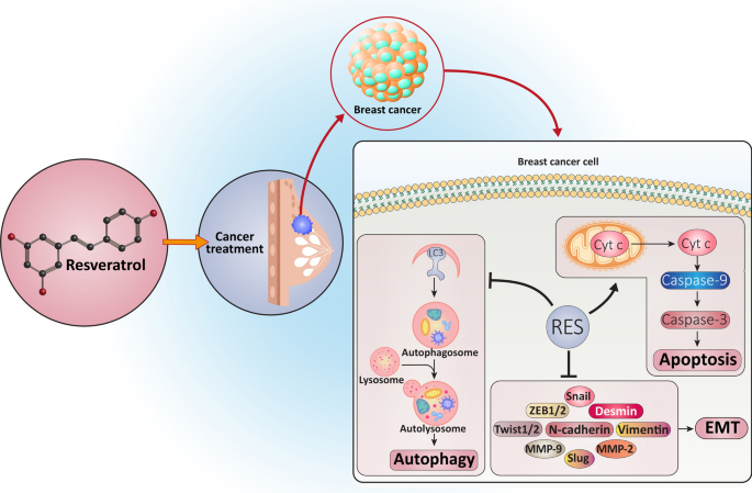
Another category of hormonal therapies includes SEEMS ( Selective Estrogen Enzyme Modulators, such as Aromatase Inhibitors) including letrozole and Arimidex, which block the conversion of androgens to estrogens in the tissue by inhibiting the aromatase enzyme. SEEMS are primarily recommended to post-menopausal women.  While most studies on resveratrol and its impact on cancer metabolism are murine or cell studies, the evidence is compelling.   Most human studies have been focused on the cell-protective, anti-aging, anti-oxidant and anti-inflammatory properties of resveratrol. Here we take a look at the aromatase inhibition properties of resveratrol.

Resveratrol (3,5,4′-trihydroxy-trans-stilbene) is a phytochemical that belongs to the stilbenoids group of phytophenolsIt is a natural plant compound found in the skin of red grapes, red wine, red grape juice, Japanese Knotweed (Polygonum cuspidatum), and in small amounts in some berries such as blueberries, mulberries, lingonberries, bilberries, red currants, cranberries and in small amounts in pistachios and peanuts. 

In one study both resveratrol, as well as melatonin, were found to be aromatase inhibitors and to have an inhibitory effect equivalent to letrozole, a commonly prescribed aromatase inhibitor drug.

Although this was true in cell culture, it has not been seen in human studies, primarily due to the fact that resveratrol has a low absorption rate when taken orally.   

Estrogen receptor-positive Anti-aromatase effect of resveratrol and melatonin on hormonal positive breast cancer cells co-cultured with breast adipose fibroblasts.


Another cell culture study demonstrated that resveratrol could inhibit aromatase at both the enzyme and mRNA expression levels and that there was a significant transcriptional control of the CYP19 gene which promotes cell proliferation in breast tissue. The research concluded that these phytochemicals can be used to target allosteric binding sites on the aromatase enzyme.

A more recent study demonstrated the anti-proliferative effect on Estrogen Receptor positive (ER+)  breast cancer cells by effectively targeting allosteric binding sites on the aromatase enzyme by resveratrol, chrysin, and apigenin. The study also included berberine and pomiferin as promising phytochemical aromatase inhibitors.  High-quality berberine is readily available and has a broad range of therapeutic actions including impacting over 20 pathways involved in cancer cell metabolism, inflammation, and bacterial pathogen control.  

Wang Y, Lee KW, Chan FL, Chen S, Leung LK. The red wine polyphenol resveratrol displays bilevel inhibition on aromatase in breast cancer cells. Toxicol Sci. 2006 Jul;92(1):71-7. doi: 10.1093/toxsci/kfj190. Epub 2006 Apr 11. PMID: 16611627.

A similar study showed that resveratrol can inhibit the CYP19 promoter gene via transcriptional control by reducing estradiol mRNA abundance through aromatase inhibition.  This leads to an anti-proliferative effect.

Wang Y, Ye L, Leung LK. A positive feedback pathway of estrogen biosynthesis in breast cancer cells is contained by resveratrol. Toxicology. 2008 Jun 27;248(2-3):130-5. doi: 10.1016/j.tox.2008.03.017. Epub 2008 Mar 29. PMID: 18462857.

The research on the impact of resveratrol on estrogen receptor-positive breast cancer proliferation and aromatase inhibition has fueled the development of several “enhanced” more bioactive resveratrol steroid analogues which may be more effective and have greater absorption than the form found in nature. 

In my clinical experience, resveratrol is a weak aromatase inhibitor and does not offer the level of protection provided by pharmaceuticals, but is also well-tolerated and without many of the adverse effects of aromatase inhibitors drugs. (Joint pain, fatigue, sleep disruption, vaginal dryness, hot flashes).  I will give 3-5 grams of pure resveratrol powder daily in 2 divided doses. To overcome the low absorption rate of resveratrol.  I always give it a high-fat food such as nut butter or full-fat yogurt and mix in cinnamon and ginger not only to improve the taste but also to enhance digestion and absorption.  This may be a good option for women who stop taking aromatase inhibitors due to unacceptable side effects negatively impacting their Quality of Life. Pharmacologic dosing of resveratrol can offer a mild aromatase inhibitory protective effect to these women along with over 50 additional pathways contributing to health and longevity.

Alhadrami HA, Sayed AM, Melebari SA, Khogeer AA, Abdulaal WH, Al-Fageeh MB, Algahtani M, Rateb ME. Targeting allosteric sites of human aromatase: a comprehensive in-silico and in-vitro workflow to find potential plant-based anti-breast cancer therapeutics. J Enzyme Inhib Med Chem. 2021 Dec;36(1):1334-1345. doi: 10.1080/14756366.2021.1937145. PMID: 34139914; PMCID: PMC8759730.

Kang H., Xiao X., Huang C., Yuan Y., Tang D., Dai X., Zeng X. Potent aromatase inhibitors and molecular mechanism of inhibitory action. Eur. J. Med. Chem. 2018;143:426–437. doi: 10.1016/j.ejmech.2017.11.057. - DOI - PubMed

Sainsbury R. The development of endocrine therapy for women with breast cancer. Cancer Treat. Rev. 2013;39:507–517. doi: 10.1016/j.ctrv.2012.07.006. - DOI - PubMed

Zhao H., Zhou L., Shangguan A.J., Bulun S.E. Aromatase expression and regulation in breast and endometrial cancer. J. Mol. Endocrinol. 2016;57:R19–R33. doi: 10.1530/JME-15-0310. - DOI - PMC - PubMed

Biomarker Lactic Acid Dehydrogenase Predicts Cancer Progression and Overall Survival

Aberrant metabolism and inefficient fuel production are characteristic of tumor cells, which are dominated by aerobic glycolysis, increased lactate production, and a higher uptake of glucose (the Warburg effect). Elevated LDH is a marker of these aberrant metabolic processes in cancer cells. High serum LDH levels are associated with poor prognosis in patients with cancer and predict progression and overall survival.

Aerobic glycolysis was described for the first time about a century ago by Otto H. Warburg who showed that cancer cells metabolize glucose differently than normal cells (Warburg effect) and that tumors derive energy mainly from the conversion of glucose to lactic acid and minimally via cellular respiration involving oxygen. Tumors produce massive amounts of the aerobic glycolysis waste product, lactic acid. This is evidence of deregulated metabolism, hence the understanding of cancer as “disorder of cellular metabolism”.  Lactic Acid itself may promote the growth and spread of cancer cells, especially at high concentrations by changing the tumor microenvironment.

Lactate dehydrogenase (LDH) is an enzyme that catalyzes the reduction of pyruvate to lactate at the end of the glycolytic pathway.

The normal range for LDH is 100-333 u/L, with levels greater than 245 u/L considered to be in the upper quartile of normal.  Elevated LDH, above 245 u/L, is suggestive of early carcinogenesis, tumor cell proliferation, tumor progression, and poor prognosis.

LDH is often highly elevated in aggressive forms of cancer and hematological malignancies including melanoma, lymphoma, acute leukemia, seminoma germ cell, pancreatic, gastric, lung, renal cell, nasopharyngeal, esophageal, cervical, and prostate cancers.

The OutSmart Cancer System® recognizes cancer as a metabolic syndrome and leverages the abnormal metabolism of tumor cells to exert influence over the tumor microenvironment and the behavior of tumor cells. Attending to the Cancer Terrain is a fundamental approach for influencing cancer cell metabolism.  

EGCG, a catechin found in Green Tea (H. Camellia sinensis) has been identified as an agent which inhibits LDH activity in normal and low oxygen environments by influencing the conversion of pyruvate to lactate at the end of the glycolytic pathway.  This may deprive cancer cells of their preferred fuel, glucose, and metabolites, including lactate that produces a favorable environment for malignant proliferation, growth, and progression. Recommended Therapeutic Dose 1-3 grams daily.

Monitoring trends in LDH is a method of both identifying abnormal cellular metabolism found in many solid and hematologic malignancies and is also of value in identifying early signs of recurrence as well as disease progression.

For patients achieving remission, during the first two years after completion of cancer treatment, LDH and other biomarkers of the Cancer Terrain are monitored every 3 months.  Thereafter, every six months for 3-10 more years to track and identify early signs of recurrence.  

For patients living with cancer as a chronic illness, LDH and biomarkers of the Cancer Terrain are monitored every 3 months to track evidence of recurrence and treatment resistance.


Learn more about monitoring the Cancer Terrain and the Tumor Microenvironment.
Receive training in Dr. Nalini’s OUTSMART CANCER SYSTEM ®.
www.aiiore.com

By using biomarkers of the Cancer Terrain and cellular metabolism, it is possible to identify trends that allow for early intervention. LDH is one of the most valuable and reliable biomarkers reflecting the active presence of the aberrant physiology of tumor cells and is prognostic and predictive of progression and overall survival in cancer patients.


Selected References:

The Connection Between Breast Cancer and The Environment

Breast Cancer is the most commonly diagnosed malignancy in women.

Image Credit - Ribbon vector created by pikisuperstar - www.freepik.com

There is a continually expanding and compelling volume of data linking breast cancer to exposure to environmental toxins, radiation and endocrine disrupters lead to increased incidence of breast cancers.

When taking a thorough history of our patients we must include a review of their “Exposome”

Genetic and Genomic factors, Reproductive history, lifestyle factors such as weight, alcohol consumption, smoking and lack of physical exercise all contribute to increased risk profiles. Socioeconomic status as well as psychological health and resilience, all influence outcomes. Racial and ethnic minorities are often exposed to a disproportionately higher level of environmental toxins in the US. Immigrants may have lived in areas where there are no environmental regulations or controls.

Exposures to common chemicals found in products used every day contribute to a lifetime burden of toxic chemicals. The greatest rise in the incidence of breast cancers occurred in the decades after World War II when there were exponential increases in the use of herbicides, pesticides, plastics, cosmetics and body care products.

Cancer is often a perfect storm of genetics and environment. While studies are done on single agents, the reality is that we are living in a toxic chemical soup in modern life exposing us to a myriad of chemicals from multiple sources on a daily basis.

A common chemical BPA (Bisphenol A) is an endocrine disruptor. Exposure to BPA early in life contributes to breast displasias later in life due to its impact on mammary gland gene expression. BPA is found in plastics, linings of canned food containers and credit card receipts.

Limit exposure to plastics, polycarbonate food and water containers and canned foods to reduce BPA exposures. Breastfeeding women should be cautious as BPA is found in human breast milk.

Parabens, p-hydroxybenzoic acid esters, are widely used preservatives in personal care products and cosmetics. Parabens are endocrine disruptors. Parabens enable the Hallmarks of Cancer, characteristics of tumor cell survival and proliferation through multiple pathways. Parabens are also found in human breast milk. Parabens bind to estrogen receptors, inhibit apoptosis, promote proliferation, angiogenesis and metastasis. A lifelong commitment to avoiding all products that contain parabens will dramatically reduce exposures. Many European countries have banned the use of parabens. European made products are often paraben free as well as select brands made in the US.

Visit the Environmental Working Group Cosmetics Data base https://www.ewg.org/skindeep/ for a list of safe and not so safe products.

Single Nucleotide Polymorphisms in P450 enzymes, particularly CYP1BI metabolism. Mulitple methylation pathways also influence detoxificaton pathways and estrogen metabolism.

A healthy microbiome, particularly rich in Bifidobacteria and butyrate support normal estrogen conjugation and excretion. MANY breast cancer treatments contribute to dysbiosis, increased inflammation and alterations in estrogen metabolism and mood.

Pelvic and Abdominal radiotherapy, surgeries, chemotherapy agents, steroids, antibiotics administered to cancer patients and compromise gut health, immunity and inflammation control. Increasing butryate in the intestines improves the health of the microbiome.

Butyrate and the health of intestinal microbiome can be easily increased by ingesting 6-8 grams of soluble fibers daily. The Onion-Garlic family and the Brassica-Cabblage family vegetables are high in soluble fibers.

The use of oral contraceptives, fertility drugs and hormone replacement therapy all alter breast tissue. Thus, medical care itself leads to nosocomial trends in breast cancer. Patients BEWARE!!!

Many pesticides and herbicides cause endocrine disruption. Commercial production of many animal food sources including the additional of estrogens and growth hormones to feed.

Patients should be well versed and take a tour of their home room by room to identify toxic, endocrine disrupting chemical exposures.

Patients can be overwhelmed when we give them a long list of products and foods to avoid.

In our clinic we employ nutritional health coaches to assist patients in successfully implementing a lifestyle and diet that reduces exposures to estrogenic environmental chemicals.

Download your complimentary copy of the
OUTSMART CANCER CARE PLANNER History and Intake Form

OutSmart Cancer Care Planner

Let The Oncologist Be The Disease Expert. Become The Health Expert That Cancer Patients Are Looking For.

You may not treat cancer in your practice, but you do have patients who are at risk due to personal and family history, patients who may be undergoing or recovering from treatments, patients who are survivors worried about recurrence and patients living with cancer as a chronic illness.  And you may also have patients who are family members concerned about their loved ones. 


There is no HEALTH MODEL in conventional oncology care, yet health and wellbeing, peace of mind and sense of agency are in the center of the hearts and minds of cancer patients, cancer survivors and their families. 


There will be 19 million cancer survivors in the US alone by 2024.  Who is supporting their health?  Who is trained to help them recover and keep them well??  …not the oncologist.


How can you help these patients?

A  breast cancer survivor who successfully completed her treatments 8 years ago comes into your office as a new patient complaining of persistent peripheral neuropathy and ongoing cognitive changes since her treatment.  How can you resolve these long-term adverse effects?


An ovarian cancer patient currently undergoing aggressive treatment every 21 days comes into your office complaining of severe diarrhea, neuropathy and sleep disruption.  What can you do to help her get through her treatments with less adverse effects, maintain her weight and nutritional status?


A colorectal cancer survivor who completed his treatment 3 months ago is continuing to have 10-15 bowel movements daily and is profoundly fatigued.  What will you do to restore normal bowel function?


A prostate cancer patient on endocrine blockade therapy is suffering from
hot flashes. Should you also be concerned about loss of bone mass and sleep cycle disruption?


An endometrial cancer survivor is suffering from dermatitis and colitis, adverse effects of her dramatically successful immunotherapy treatment and now has chronic autoimmune inflammation. How will you manage this?


A head and neck cancer patient who has trouble swallowing is losing weight and muscle mass.


How can you provide a plan for repair from oral mucositis, restoration of the oral mircrobiome and repletion of calories and nutrients?


These patients are searching for clinicians that can guide and support them through every phase of their cancer journey.  Just as in helping your patients navigate other chronic illnesses, patients look to you for a plan, for monitoring and guidance so that they can maintain and regain their health during and after their treatments.


When a patient has a collaborative team providing integrative care everyone wins, the patients, families and care providers.  Patients who have a clear plan and support have the opportunity for better outcomes, better prognosis, greater peace of mind, a sense of control and agency and an improved quality of life. 


Let the oncologist be the cancer expert. You can be the health expert on their team.


Standard of care in oncology must  change such that care includes not only a team of disease experts (usually medical oncologist, surgeon, radiologist) but ALSO a team of health experts.


Towards this end  I founded the American Institute of Integrative Oncology Research and Education and  have created an online self-paced training program for front line clinicians who want to expand their skills and their practice and  fill the huge need in our communities and serve these patients.  If you did not specialize in oncology, you probably had one course on this topic but you need to fill the gap in your training to feel confident in doing so.


The Foundations of Integrative Oncology Training is not for clinicians who want to practice oncology.  It is front-line clinicians who want to feel confident, knowledgeable and well trained in supporting the health side of the cancer equation. This self- paced online training is for clinicians who want to increase their impact, expand and grow their practice and represents 35 years of clinical practice and experience.


The first step is learning how to take a comprehensive and complete history of patients whose lives have been touched by cancer.  


You can receive a complimentary copy of the

OUTSMART CANCER CARE PLANNER History and Intake Form

and learn more about the Foundations of Integrative Oncology training here


OutSmart Cancer Care Planner