Home » OutSmart Cancer » Page 2

Tag: OutSmart Cancer

cancer-patient

VITAL TALK: Learn to Communicate Effectively with Patients Experiencing Serious Illness

“Just as no doctor is born knowing how to handle a scalpel, the same is true for how to communicate effectively with seriously ill patients and their families. We believe every clinician can become a better communicator.”

dr-tony

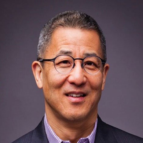
I met oncologist Dr. Tony Back, MD at a meditation retreat focused on working skillfully and mindfully with patients with serious, life changing illnesses as well as end of life care.  Dr. Back is affiliated with the Center to Advance Palliative Care at Fred Hutchinson Cancer Center in Seattle, Washington and has combined his years of clinical oncology experience with his recognition that patients and clinicians require more skillful and effective conversations in the midst of emotionally charged and medically complex discussions. Dr. Back has developed online and in person trainings called Vital Talk for clinicians to LEARN TO COMMUNICATE EFFECTIVELY WITH PATIENTS EXPERIENCING SERIOUS ILLNESS. His mission is “To Elevate Patient Care with better communication”.

Dr. Back and the Vital Talk mission is “that every seriously ill patient will be surrounded by clinicians who can speak about what matters most and match care to values.”

Vital Talk Trainings help clinicians to Master Tough Conversations

Whether in person or online, clinicians feel safe practicing newly learned skills through VitalTalk’s evidence-based training methodologies.

Some tips from a Vital Talk trained physician

Dr. Nalini

  • Give yourself grace
  • Lean in with emotion, heart and feeling
  • Respond with empathy.  
  • Explore and ask the patient to tell you more.  
  • Ask permission to talk about a topic (scan results, disease progression, end of life care)

[READ MORE]

EVIDENCE-BASED SKILLS Vital Talk TRAINING COURSES 

  • Elevate patient care with better communication
  • Explore best-practice communication methodologies and tools, as well as our rich community of support to better serve the needs of patients with serious illness and their families.
  • Support clinicians in learning to communicate effectively and compassionately with patients living with serious illness. 

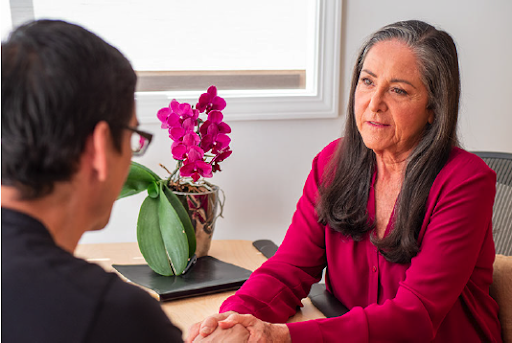
We are living in the midst of an aging patient population in the United States. Therefore our patients are increasingly at risk of developing more serious illnesses, experiencing co-morbidities and facing many life changes of loss and limitation, frailty and mortality.   Cancer patients of all ages face immense challenges and must redefine their sense of self and examine their values and relationship to life, suffering, death and dying. These challenges may be turned into gateways to transformation.  I counsel my patients to reframe their cancer journey so that the challenges that they face become opportunities for growth, insight and wisdom and not just suffering, grief and loss.   These conversations require clinicians to develop greater communications skills and the ability to be comfortable engaging in these dialogues.

It is my experience that clinicians who are inspired to work with cancer patients are motivated by the drive towards making a difference, the intellectual challenge of the complexity of cancer and the deep meaning that arises in meeting patients at the edge where  the rawness and overwhelm of being a cancer patient can be transformed into a profound healing journey of awareness and transformation.   

I highly recommend that all clinicians continue to develop their clinical skills so that they can be fully present, capable and comfortable for participating in these most tender, human and meaningful conversations.   

I always feel it is a privilege and an honor to be allowed to accompany a patient and their family on their journey and to be able to guide them into deeper meaning and a greater sense of connection to each other and to all of life.

I encourage you to explore The Vital Talk website where you will find information on their programs and trainings but also Free Downloadable Tools and Guides to get you started

https://www.chooseyourpath.vitaltalk.org/

https://www.vitaltalk.org/resources/

Please do share your stories with us!!  

Cancer-SARS

Inflammation, Cancer and SARS-CoV2

Managing Inflammation and Inflammasome in both the Cancer Terrain and SARS-Cov2

There is a subset of cancer patients who suffer significantly more inflammation as well as the sequela of increased inflammation including ongoing cancer related fatigue, increased pain, cognitive deficits.   Similarly there is a subset of COVID-19 patients who suffer a cytokine storm and the wildfire of inflammation that leads to respiratory distress syndrome and mortality. Identifying patients with a higher risk of increased inflammation can be assessed by taking a thorough history in search of historical tendencies and patterns and an analysis of single nucleotide polymorphisms (SNPs).  Patients with IL1B, IL6 and NFkB  SNPs are more prone to developing greater inflammation in both syndromes.

inflammationThe inflammatory drivers and cytokines are similar in both cases: NFkB, TNFa, IL1, IL6, IL8, Inflammasone NLRP3, TGFb1, STAT3, JAK2, p38MAPK, Nrf2, AMPK

Activation of Inflammasome NLRP3 is correlated with the development of the SARS CoV2 cytokine storm.  Inflammasome activation is an important component of innate immunity which enhances inflammation.  Inflammasome activity is correlated with destructive inflammation particularly in viral diseases.  Inflammsome NLRP3  increases IL-1B is also  upregulated in the cancer terrain, particularly in metastatic lung cancer, breast cancer, fibrosarcoma and gastric carcinoma.

curcuminCurcumin exhibits anti-inflammatory and anti-inflammasome properties and can be used in both syndromes.  Curcumin impacts all of the above named inflammatory drivers along with inhibition of COX2 transcription.    Furthermore curcumin acts as an antioxidant increasing control of reactive oxygen species present with increased inflammation.

I recommend Designs for Health Curcumevail, Thorne Research Meriva and Euromedica Curapro.  In managing both the cancer terrain and the SARS CoV2 terrain the dose range is 2g-6g curcuminoids per day.

Another actor, Nrf2, is a nuclear transcription factor that increases the presence of antioxidant proteins when cells are stressed.   Management of the cancer terrain also includes support for normal function of Nrf2.  Nrf2 is highly expressed in the lungs and is responsible for inhibiting the activity of Inflammasome NLRP3.  Sulforaphanes include Di-indole methane, Indole-3-Carbinol and  Sulforaphane glucosinolate.  Broccoli, broccoli sprouts and kale are dietary sources of sulforaphanes.  

antioxidant

I recommend Designs for Health Broccoprotect and Thorne Research Crucera for a high quality source of sulforaphane glucosinolate 50mg twice daily.

Ultimately we may find that the core foundation and targeted supplements used in the OutSmart Cancer System for managing the cancer terrain ALSO protect our patients in the midst of a viral pandemic.

Selected References 

Rajendra Karki et al Inflammasomes and Cancer                                                                                                             PMID: 28093447 DOI: 10.1158/2326-6066.CIR-16-0269

Saeedi‐Boroujeni  et al COVID‐19: A Case for Inhibiting NLRP3 Inflammasome, Suppression of Inflammation with Curcumin?  Ali  https://doi.org/10.1111/bcpt.13503 Volume128, Issue1 January 2021 Pages 37-45

Howrylak JA, Nakahira K. 

Inflammasomes: Key Mediators of Lung Immunity. 

Annu Rev Physiol. 2017;79:471‐494. doi:10.1146/annurev-physiol-021115-105229

James W.Pinkertona1Richard Y.Kima1Avril A.B.RobertsonbJeremy A.HirotacLisa G.WoodaDarryl A.KnightaMatthew A.CooperbLuke A.J.O’NeilldJay C.Horvata1Philip M.Hansbroa  Inflammasomes in the Lung

Molecular Immunology Volume 86, June 2017, Pages 44-55         

Shih-Yi Chuang,1,2 Chih-Hung Lin,3,4 and Jia-You Fang

Natural Compounds and Aging: Between Autophagy and Inflammasome

Biomed Research Int. Volume 2014 |Article ID 297293 | 10 pages | https://doi.org/10.1155/2014/297293                                                    

József Tőzsér1 and Szilvia Benkő

Natural Compounds as Regulators of NLRP3 Inflammasome-Mediated IL-1β Production

Mediators of Inflammation Volume 2016 |Article ID 5460302 | 16 pages                                               https://doi.org/10.1155/2016/5460302

turmeric

Tumeric Prevents Chemotherapy Induced Hand Foot Syndrome

Xeloda (Capecitabine) is the oral form of  chemotherapy agent 5-Fluorouracil  (5-FU) which is administered intravenously. 

Both drugs are widely used in a broad range of cancers. The mechanism of action is inhibition of mitosis (cell division).   

One of the most common side effects of these drugs is HAND-FOOT SYNDROME or palmar-plantar erythrodysesthesia.  Hand-Foot syndrome presents with redness, swelling and pain, tingling, burning and sensitivity to touch on the palms of the hands and the soles of the feet. Severe cases may include cracked, flaking or peeling skin, painful blisters, ulcers or sores, severe pain and difficulty walking or using the hands.

While etiology is not certain it is hypothesized that these drugs cause capillary fragility and subsequent leakage of drugs into and damage of  the surrounding tissue.

fhs

Other commonly used chemotherapy agents  which result in Hand - Foot syndrome include 5-Fluorouracil, Capecitabine, Cytarabine, Docetaxel, Doxorubicin, Doxil and Paclitaxel. These drugs are widely used in Breast, Ovarian and Gastrointestinal Cancers.  

Not all patients who receive these drugs develop Hand-Foot Syndrome. The symptoms typically appear with the first dose administration in 40-50 percent of patients at grade 2 or higher.

turmeric-as-cure

Grade 1: painless erythema or dysesthesia, no impairment

Grade 2: painful erythema, swelling, tingling, numbness, dryness, cracking, Desquamation, activity is impaired

Grade 3: strong pain, ulceration, blistering, erythema, limited self-sufficiency

For drugs administered via IV infusion, cold (Ice) gloves and booties that constrict capillaries reduce local exposures in the hands and feet and must be worn during infusions and for several therapy to hands and feet concurrently with chemotherapy infusions. 

Capcetabine increases COX-2 expression in both tumors and healthy tissues.

A double blind placebo controlled study in which oral COX2 inhibitor celecoxib was administered to patients over a 2 year period while receiving capecitabine or capecitabine plus oxaliplatin demonstrated a significant decrease in the incidence and intensity of symptoms.

The phytochemical curcumin derived from Rhizoma Curcuma longa interacts with over 100 genes that impact tumor cell behavior and metabolism. Curcumin is known to decrease COX2 expression along with expression of  pro-inflammatory NFkB, TNFa, IL1B, IL6 and IL8.  

foot-hand-syndromeIn a human study researchers administered turmeric at a dose of 4 g/day (2 pills 12 hours apart) starting at the beginning of capecitabine treatment and lasting six weeks. The study included 40 patients whose mean age was 62 years. Most were female (80%), 52% had breast cancer, and 47.5% had GI tumors. After the first cycle of capecitabine treatment, 11 of 40 patients developed HFS (27.5%; 95% CI [15, 42]), whereas four patients developed HFS equal or superior to grade 2 (10%; 95% CI [3.3, 23]).. The study concluded that  turmeric combined with capecitabine seems to produce a lower rate of HFS, especially grade 2 or higher. 

Comments

  • There were no contra-indications to utilizing turmeric concurrently with capecitabine
  • A significant therapeutic dose of 4 grams per day was used.   In my practice I prefer to use the more concentrated isolate curcumin and dose at 2-4 grams per day.  This would yield more inflammation control.   
  • Turmeric falls into the category of herbs that “Move Stagnant Blood” in Chinese Medicine.  One of the properties of curcumin is inhibition of platelet aggregation.

References:

Curcuma longa (Turmeric) for Prevention of Capecitabine-Induced Hand-Foot Syndrome: A Pilot Study

Vanessa Armenio Scontre , MD,Janine Capobiango Martins , MD, et al 

Jnl Diet Supp Pages 606-612 | Published online: 02 Nov 2017

 https://doi.org/10.1080/19390211.2017.1366387 PMID: 2909565

The effect of COX-2 inhibitor on capecitabine-induced hand-foot syndrome in patients with stage II/III colorectal cancer: a phase II randomized prospective study.Zhang RX, Wu XJ, Lu SX, Pan ZZ, Wan DS, Chen G.
J Cancer Res Clin Oncol. 2011 Jun;137(6):953-7. doi: 10.1007/s00432-010-0958-9. Epub 2010 Nov 27.
PMID: 21113620 Clinical Trial.

stress-cancer

Does Stress Cause Cancer?


Lifestyle Factors That Impact Breast Cancer Risk

cancer-and-stress

  • Alcohol:  Drinking Alcohol Increases Risk of Breast Cancer
  • Weight and Body Composition: Excess body fat increases risk for post-menopausal breast cancer. Lean muscle, low body fat decreases risk of pre-menopausal breast cancers
  • Physical Activity: Sedentary behavior is linked to increased risk of breast cancer, while being active decreases the risk of breast cancer 

Vigorous activity decreases the risk for pre-menopausal breast cancer.

Moderate activity decreases risk for post-menopausal breast cancer.

Some evidence indicates that people who are physically active (both before and after diagnosis) have a greater chance of surviving breast cancer.

  • Breastfeeding: Reduces risk of both pre- and post-menopausal breast cancer
  • Sleep: Women who report sleeping less than 5 hours per night  before diagnosis have an increased risk of dying from breast cancer compared to women whose  pre-diagnosis sleep pattern was 7-8 hours per night.  Women who have disrupted circadian rhythms due to night shift work have an increased risk of breast cancer.

cancer-cells

Does Stress Cause Cancer?

Maladaptive and ongoing responses to stress mediated by the Autonomic Nervous System and Hypothalamic Pituitary Axis promote a tumor microenvironment that favors inflammation, oxidative stress, poor glycemic control, carcinogenesis, proliferation, angiogenesis and metastasis

Physiological Pathways, Bio-behavioural Processes and Oncogenesis:

  • Environmental and social processes activate interpretive processes in the central nervous system (CNS) that can subsequently trigger fight-or-flight stress responses in the autonomic nervous system (ANS) or defeat/withdrawal responses through the activation of the hypothalamic–pituitary–adrenal axis (HPA)
  • Individual differences in perception and evaluation of external events (coping) creates variability in individual ANS and HPA activity levels.
  • Over long periods of time, these neuroendocrine dynamics can alter various physiological processes involved in tumorigenesis, including oxidative metabolism, DNA repair, oncogene expression by viruses and somatic cells, and production of growth factors and other regulators of cell growth.
  • Once a tumour is initiated, neuroendocrine factors can also regulate the activity of proteases, angiogenic factors, chemokines and adhesion molecules involved in invasion, metastasis and other aspects of tumour progression.
  • CNS processes can also shape behavioural processes that govern cancer risk (for example, smoking, transmission of oncogenic viruses or exposure to genotoxic compounds).


Integrated Model of Bio-behavioral Influences on Cancer Pathogenesis Through Neuro-Endocrine Pathways

chart

In this model, bio-behavioural factors such as life stress, psychological processes and health behaviours (blue panel) influence tumour-related processes (green panel) through the neuroendocrine regulation of hormones, including adrenaline, noradrenaline and glucocorticoids (red panel). 

Central control of peripheral endocrine function also allows social, environmental and behavioural processes to interact with biological risk factors such as genetic background, carcinogens and viral infections to systemically modulate malignant potential (red panel). 

Direct pathways of influence include effects of catecholamines and glucocorticoids on tumour-cell expression of genes that control cell proliferation, invasion, angiogenesis, metastasis and immune evasion (green panel). 

Stress-responsive neuroendocrine mediators can also influence malignant potential indirectly through their effects on oncogenic viruses and the cellular immune system (red panel). 

These pleiotropic hormonal influences induce a mutually reinforcing system of cellular signals that collectively support the initiation and progression of malignant cell growth (green panel). 

Furthermore, neuroendocrine deregulation can influence the response to conventional therapies such as surgery, chemotherapy and immunotherapy (green panel). 

In addition to explaining bio-behavioural risk factors for cancer, this model suggests novel targets for pharmacological or behavioural intervention. 

(CTL, cytotoxic T lymphocytes; IL, interleukin; MRD, minimal residual disease; NKC, natural killer cell; TGFβ, transforming growth factor-β; TNFα, tumour-necrosis factor-α; TSH, thyroid-stimulating hormone.)

Dr. Nalini’s Adrenal Stress-Immune Support Protocol 

DAYTIME

Designs for Health Adrenotone   2/3x/day. With meals

All-in-one synergistic adrenal support formula. 

Metagenics Immucore 1/3x/day. With meals

Multidimensional Support for Healthy Immune Function

BEDTIME

Integrative Therapeutics  Cortisol Manager 2 caps one hour  before bedtime

  • Safe for use every night
  • Stress reducing sleep aid
  • Reduces cortisol levels for stress reduction and restful sleep.

https://us.fullscript.com/protocols/chilkov-dr-nalin-s-adrenal-stress-immune-support-kit

Treatment Plan should include 

Patient Teaching, Lifestyle and Dietary Guidelines and ongoing behavior change support

  • Dietary Guidelines -Nutrient Repletion-Glycemic Control
  • How to nurture parasympathetic tone
  • Sleep Hygeine
  • Self Regulation-Resilience- Stress and Mood Management guidelines
  • Monitoring Heart Rate Variability
  • Encourage Meditation-Tai Chi-Yoga-Deep Relaxation, Time in Nature
  • Acupuncture
  • Massage
  • Importance of Social Support
green tea extracts

Green Tea Extract Reduces Severity of Radiation-Induced Dermatitis

Breast cancer is the most common cancer affecting women worldwide. Radiation dermatitis affects nearly all women receiving radiotherapy for breast cancer.  http://repoceuticals.dk/pipeline/radiation-dermatitis/

new-breast-cancer

RID may result in less tolerance to treatment and even discontinuation of treatment. The patient may have skin changes ranging from faint erythema (reddening) and desquamation (peeling skin) to skin necrosis (death of skin cells) and ulceration, depending on the severity of the reaction.  Several studies demonstrate that topical green tea extract may be an effective prophylactic treatment.  

To make a strong hot water extract:  Place 8 tea bags of organic green tea into 1 cup of filtered or distilled water. Bring to a boil and simmer covered for 20 minutes. Place extract into sterile glass dropper bottle. (Available at most pharmacies).   Spray skin liberally before and after radiotherapy treatment.

The National Cancer Institute (USA) has developed 4 criteria for the classification of acute radiation dermatitis:

  • Grade 1 – Faint erythema or desquamation.
  • Grade 2 – Moderate to brisk erythema or patchy, moist desquamation confined to skin folds and creases. Moderate swelling.
  • Grade 3 – Confluent, moist desquamation greater than 1.5 cm diameter, which is not confined to the skin folds. Pitting oedema (severe swelling).
  • Grade 4 – Skin necrosis or ulceration of full-thickness dermis (middle layer of skin).

Hymes SR, Strom EA, Fife C. Radiation dermatitis: Clinical presentation, pathophysiology and treatment 2006. J Am Acad Dermatol 2006; 54:28-46. PubMed 

https://dermnetnz.org/topics/radiation-dermatitis

Epigallocatechin-3-gallate ameliorates radiation-induced acute skin damage in breast cancer patients undergoing adjuvant radiotherapy

In a study using topical Epigallocatechin-3-gallate forty nine patients topical EGCG was applied daily during radiotherapy treatment.  “Topical EGCG was applied daily, starting when grade I dermatitis appeared and ending two weeks after radiotherapy. The maximum dermatitis observed during the EGCG treatment was as follows: Grade 1 toxicity, 71.4% (35 patients); grade 2 toxicity, 28.6% (14 patients); there were no patients with grade 3 or 4 toxicity. The majority of the radiation-induced dermatitis was observed 1 week after the end of radiotherapy. EGCG reduced the pain in 85.7% of patients, burning-feeling in 89.8%, itching in 87.8%, pulling in 71.4%, and tenderness in 79.6%.”

Zhu W et al Oncotarget. 2016 Jul 26;7(30):48607-48613. doi: 10.18632/oncotarget.9495. PMID: 27224910; PMCID: PMC5217042.

Efficacy of Epigallocatechin-3-Gallate in Preventing Dermatitis in Patients With Breast Cancer Receiving Postoperative Radiotherapy: A Double-Blind, Placebo-Controlled, Phase 2 Randomized Clinical Trial 

A total of 180 eligible patients were enrolled, of whom 165 (EGCG, n = 111; placebo, n = 54) were evaluable for efficacy (median [range] age, 46 [26-67] years). The occurrence of grade 2 or worse RID was significantly lower (50.5%; 95% CI, 41.2%-59.8%) in the EGCG group than in the placebo group (72.2%; 95% CI, 60.3%-84.1%) (P = .008). The mean RIDI in the EGCG group was significantly lower than that in the placebo group. Furthermore, symptom indexes were significantly lower in patients receiving EGCG. Four patients (3.6%) had adverse events related to the EGCG treatment, including grade 1 pricking skin sensation (3 [2.7%]) and pruritus (1 [0.9%]).

Prophylactic use of EGCG solution significantly reduced the incidence and severity of RID in patients receiving adjuvant radiotherapy for breast cancer.

Zhao H, Zhu W,  et al. JAMA Dermatol. 2022 Jul 1;158(7):779-786. doi: 10.1001/jamadermatol.2022.1736. PMID: 35648426; PMCID: PMC9161122.

tea-extracts

The effects of tea extracts on proinflammatory signaling

Tea extracts supported the restitution of skin integrity. 

Tea extracts inhibited proteasome function and suppressed cytokine release. 

NF-kappaB activity was altered by tea extracts in a complex, caspase-dependent manner, which differed from the effects of epigallocatechin-gallate. 

Additionally, both tea extracts, as well as epigallocatechin-gallate, slightly protected macrophages from ionizing radiation

Pajonk F, et al, BMC Med. 2006 Dec 1;4:28. doi: 10.1186/1741-7015-4-28. PMID: 17140430; PMCID: PMC1698929.

doi: 10.1186/1741-7015-4-28. PMID: 17140430; PMCID: PMC1698929.

JAMA Derma

environmental-toxins

All Cancers Linked to Environmental Toxins

Making Environmental Health A Part of Health Care

Each month of the year is devoted to awareness of one or more cancers. September is devoted to multiple cancers.  

What do all of these cancers have in common?

There is an increasing volume of compelling evidence linking all types of cancer to environmental exposures.

Our knowledge of mechanisms linking exposures of toxicants to specific cancers is also increasing.  

environmental-pollution

There are two reasons that we as clinicians and care providers must be aware of the many contributing offenders and how to identify, assess, mitigate risk and safely remove body burden or toxicants in our patients.

Additionally, it is my practice to engage in patient education and patient teaching on the first or second visit to increase patient and family awareness about the importance of taking control of and reducing their toxic exposures as many toxicants are ubiquitous our homes, workplaces and communities.  Many toxicants include the use of common everyday products.    I also refer patients to these very reliable and up to date websites:  Environmental Working Group where foods, cleaning supplies, garden supplies are rated and their secondary site  Skin Deep Cosmetics Database for self-care and baby care products, including safe sunscreens.  They also have many downloadable publications for patients including 12 Hormone-Altering Chemicals and How to Avoid Them

Taking a good “ toxic exposures history” is important in all patients.  Of course, the very diagnostic methods and treatment modalities used in oncology are sources of toxic exposure as well!!  Many clinicians feel taking such a history from their patients will open us a Pandora’s box.

sewer-pollution

However, in the context of oncology, it is essential to do so, if only to bring awareness and to address the risk and health status not only of the patient, but also their family members.

My go to experts in this area include Dr. Walter Crinnion ND and Joseph Pizzorno, ND, both seasoned researchers and clinicians. They have recently published a well researched book  Clinical Environmental Medicine, which I consider required reading for all clinicians, especially those working with cancer patients.  There is a version for patients as well The Toxin Solution: How Hidden Poisons in the Air, Water, Food, and Products We Use Are Destroying Our Health--AND WHAT WE CAN DO TO FIX IT Dr. Crinnion’s has an excellent downloadable Toxic Exposure Questionnaire.  

The  American Academy of Environmental Medicine is an excellent resource for education and training and conferences where world-class experts convene.

Most in the patients control is their home environment and their food and water.

Fran Drescher’s Cancer Schmancer website has instructions for how to host a Detox Your Home Party.   

The first step is avoidance, avoidance, avoidance to reduce body burden, followed by appropriate supplements to help the body deal with what is there and finally safe and appropriate cleansing and detoxification interventions. Patients must also understand how to avoid re-exposure.

Resources:

Recommended Laboratories for Assessing Body Burden of Toxins 

(This is not a complete list and I have no financial relationship with any of these labs)

  • Great Plains Laboratory (heavy metals, environmental exposures, mycotoxins, glyphosate)
  • Genova Diagnostics (heavy metals, environmental chemicals)
  • RealTime Labs (Mold and Mycotoxins)
  • IMS Laboratory (Mold and Mycotoxins)
  • Quicksilver Scientific (Mercury)

*from a headline published in the New York Times

green-tea-extract

Green Tea Extract Reduces Severity of Radiation Dermatitis

The use of a solution containing a green tea extract has been shown to reduce both the incidence and severity of radiation-induced dermatitis in women undergoing adjunctive radiotherapy for breast cancer. This was the conclusion of a phase 2 randomized, placebo-controlled trial by a team of Chinese researchers.

Data from the World Health Organization indicates that in 2020, there were 2.3 million women diagnosed with breast cancer. In the treatment of women with breast cancer, radiation therapy is widely used conjunction with other therapies such as surgery, chemotherapy and hormonal therapies.  A common and frequent adverse effect of radiotherapy is radiation-induced dermatitis (RID) suffered by millions of women.

green-tea

The purpose of the current  study was to investigate the safety, tolerability and preliminary effectiveness of topical epigallocatechin-3-gallate (EGCG) for radiation dermatitis in patients with breast cancer receiving adjuvant radiotherapy.

A  solution of green tea extract  sprayed on the radiated areas of the skin reduced severity of radiation-induced dermatitis.

The Chinese team recruited women with breast cancer undergoing postoperative radiotherapy and randomized them (2:1) to receive either the green tea extract or placebo (normal saline solution).  These solutions were sprayed to the whole of the radiation field from the first day of therapy until two weeks after completion of treatment. 

A total of 165 women with a median age of 46 years were enrolled and randomized to EGCG, the primary catechin found in green tea or placebo.

The onset of radio-dermatitis was delayed by 2-3 weeks and the intensity and severity of the symptoms were significantly decreased in the treated group.  No skin toxicity was observed.

The authors concluded that prophylactic use of a green tea extract significantly reduced both the incidence and severity of RID and that it has the potential to become a new choice for skin care in women receiving radiotherapy.

Topical green tea extract supports restoration of skin integrity and control of inflammatory cytokines and oxidative stress in the skin. Green tea extract also reduces the acute skin-induced reactions including pain and sensations of burning, itching, pulling and tenderness.

Dr. Chilkov: Practical Application:

green-tea-leavesTopical Green Tea Extract Spray

To make a medicinal water extract: Place 8 organic green tea bags into a 16 oz glass jar or glass container.  Pour boiling water over the tea bags, cover immediately and steep for one hour.   After it has cooled to room temperature store covered in the refrigerator.  When ready to use transfer water extract to a a glass spray bottle.  Apply liberally to the radiation field before and after each radiotherapy session and three times daily for 3 weeks after the last radiotherapy session.  

Fresh Aloe Vera Gel poultice

Areas where skin is most impacted can be covered with. mashed fresh aloe vera gel and covered with a large gauze bandage.   This can easily be held in place underneath a sports bra or leotard or similar.  Apply fresh aloe gel twice daily. Allow to be in contact with the skin for several hours or overnight.  If you do not have access to a live aloe vera plant or fresh aloe gel you can use alcohol free aloe vera juice or aloe vera gel commonly found in natural foods stores.    Aloe Vera is the botanical of choice for repair of radiation damaged skin.

Topical Calendula Oil (not extract) is also a soothing topical anti-inflammatory agent for radiation induced dermatitis.  If the skin is very damaged, saturate a 4x4” gauze square and place over the affected area.

References:

Zhao H et al. 

Efficacy of Epigallocatechin-3-Gallate (EGCG)in Preventing Dermatitis in Patients With Breast Cancer Receiving Postoperative Radiotherapy: A Double-Blind, Placebo-Controlled, Phase 2 Randomized Clinical Trial JAMA Dermatol 2022

Zhao H, et al. 

Phase I study of topical epigallocatechin-3-gallate (EGCG) in patients with breast cancer

receiving adjuvant radiotherapy. Br J Radiol 2016; 89: 20150665.

Kyle T. Amber, BS et al

The Use of Antioxidants in Radiotherapy-Induced Skin Toxicity 

Integrative Cancer Therapies 2014, Vol. 13(1) 38–45

Book Review: You Finished Treatment-Now What?


A Field Guide for Cancer Survivors.
By Dr. Amy Rothenberg

You-Finished-Treatment-Now What

You Finished Treatment, Now What? A Field Guide for Cancer Survivors by Dr. Amy Rothberg is a roadmap for lifestyle and natural medicine approaches to address health challenges that persist after cancer care, and to reduce the risk of recurrence.

Dr Rothenberg wrote this guide for cancer survivors and those on their support and care team.

You Finished Treatment, highlights the evidence for an integrative approach to healing that Dr. Rothenberg has used for over 37 years practicing as a licensed naturopathic doctor.

She is also a breast and ovarian cancer survivor/thriver herself. She wrote this book to make sense of an overwhelming topic, in a user-friendly, accessible way, providing both actionable information and inspiration.

As a survivor/thriver of both ovarian cancer and breast cancer she speaks both from the physician’s and the patient’s point of view with heart, levity and solid, practical advice.

When diagnosed with cancer in 2014, Dr. Rothenberg sought and received state-of-the-art care at a renowned teaching hospital and had her own naturopathic medical team to help her best handle treatment, and rebound afterward.

Her writing is evidence-informed, while also bringing her personal experience as a doctor, patient, wife, mother, sister, and friend. Offering a natural, integrative medicine perspective on items in the news, find Dr.

This is an example of the principles employed in Dr. Chilkov’s OutSmart Cancer® System which is an integrative approach to combining the best of modern oncology with the best of research informed modern and traditional naturopathic systems of medicine for the very best outcomes.

This approach allows patients to have not only a plan for their disease, but also a plan to support their health during and after treatment and to support recovery, restoration and rejuvenation in support of both healthspan and lifespan.

Dr. Rothenberg’s both personal experiences and medical expertise combine to form a heartful and pragmatic approach with clear guidelines and recommendations. This book is a wonderful resource for both patients and families as well as care providers.

Breast-Cancer

Changing the Management of Cancer with Personalized Testing


Personalized cDNA surveillance for patients with high-risk breast cancer

Is there a more sensitive technology that can detect preclinical breast cancer progression?

It is now possible to monitor fragments of cell free tumor DNA (ctDNA) circulating in the blood. This falls under the umbrella of “liquid biopsies” which monitor tumor burden, tumor response to treatment and early signs of recurrence or progression without a scan or need for a new surgical or biopsy tissue sample.

  • “Up to 30% of patients with breast cancer relapse after primary treatment.
  • There are no sensitive and reliable tests to monitor these patients and detect distant metastases before overt recurrence.
  • Breast cancer cell free tumor DNA blood test (liquid biopsy) can detect recurrence up to 2 years earlier than currently available conventional serum tumor markers and radiologic studies.
  • Cell free tumor DNA assays predict breast cancer recurrence earlier and with greater accuracy than traditional tools by using a highly-personalized molecular residual disease assay.

A cell free tumor DNA (ctDNA) assay is a personalized, tumor-informed assay with the power to give you earlier, clearer insight into your patient's disease. By detecting and quantifying ctDNA, you can optimize your ability to assess risk, predict recurrence, and monitor treatment response in those most at risk for progression.

This technology can be used to monitor a wide range of cancers. While this type of monitoring has not yet been widely adopted as “standard of care” I encourage you to educate all of your patients and their care providers to adopt the use of this highly reliable screening tool now.

Here, we demonstrate the use of personalized circulating tumor DNA (ctDNA) profiling for detection of recurrence in breast cancer.”

Cell free tumor DNA assays use a sample of the patient’s tumor tissue to develop a unique DNA fingerprint. After that, follow-up blood draws capture changes in the level of ctDNA, giving clinicians a better picture of a patient’s risk of recurrence without the need for another tissue sample and may decrease the need for frequent scans and repeated frequency of exposure to radiation and contrast material.

neodjuvant

A recent study “demonstrates that patient specific ctDNA analysis can be a sensitive and specific approach for disease surveillance for patients with breast cancer. More importantly, earlier detection of up to 2 years provides a possible window for therapeutic intervention. “(1)

Currently, there are no sensitive and specific clinical tests available to follow patients with breast cancer after primary treatment. Signatera developed a patient-specific method to analyze circulating tumor DNA (ctDNA) that allows for monitoring of these patients regardless of molecular genotype. In this study, we analyzed 208 blood samples from 49 patients monitored longitudinally for up to 4 years after completion of adjuvant chemotherapy to determine whether personalized ctDNA assays can allow for more effective monitoring than current clinical tests such as CA 15-3. Remarkably, for the patients that recurred, our test detected molecular relapse up to 2 years ahead of clinical relapse (median, 8.9 months) with 89% sensitivity and 100% specificity. This may provide a critical window of opportunity for additional therapeutic intervention.” (1)

hope

Data from a retrospective cohort analysis of EBLIS, a study designed to determine the lead interval between ctDNA detection and clinical metastatic disease, and to determine whether ctDNA in plasma can detect recurrent disease earlier than traditional methods, demonstrated that Signatera can accurately predicts metastatic relapse with a significant lead time over imaging and CA 15-3 (200 days on average)

Neoadjuvant

“…our study shows promise that early response prediction by highly sensitive ctDNA analysis in high-risk early breast cancer patients may facilitate a timely and judicious change in treatment to improve patients’ chances of achieving favorable long-term outcomes.(2)

Surveillance

Patients undergoing treatment as well as those who have completed their course of treatment can be assessed both for response to treatment during a course of therapy as well as for early signs of reurrence after treatment has been completed. In a study of patients undergoing treatment with Pembrolizumab, a checkpoint inhibitor.

“Baseline ctDNA concentration correlated with progression-free survival, overall survival, clinical response and clinical benefit. This association became stronger when considering ctDNA kinetics during treatment. All 12 patients with ctDNA clearance during treatment were alive with median 25 months follow up. This study demonstrates the potential for broad clinical utility of ctDNA-based surveillance in patients treated with ICB.” (3)

Recommended labs offering this technology include Natera, INVITAE, Foundation One, Caris Life Sciences. All of these labs are highly regarded in the oncology community. (Disclosure: I have no financial relationships with any of the labs recommended in this article.)

How often should these assays be performed?
I recommend monitoring monthly during active treatment to determine if the current treatment is effective and continuing to be effective. This is a way to identify treatment resistance early.

top10-badgeI recommend monitoring every three months during the first two years after completing treatment or for patients with advanced receiving ongoing treatments. (For example advanced breast cancer patients receiving hormonal treatments, immunotherapy treatments, checkpoint inhibitor treatments or chemotherapy treatments over long periods of time.)

For long term survivors I recommend monitoring every 6 months until the 10 year No Evidence of Disease anniversary.

This is the same schedule of monitoring that we use in the OUTSMART CANCER® System to follow measurable biomarkers in the tumor microenvironment.

Discover how you can join
Foundations of Integrative Oncology Professional Online Training
Master the OUTSMART CANCER® System
Receive Monthly Live Mentoring and Case Supervision
from Dr. Nalini Chilkov
CLICK HERE

References

  1. Coombes C, Page K, Salari R, et al. Personalized Detection of Circulating Tumor DNA Antedates Breast Cancer Metastatic Recurrence. Clinical Cancer Research. 2019;25(14):4255-4263.
  2. Circulating tumor DNA in neoadjuvant-treated breast cancer reflects response and survival M. J. M. Magbanua. https://doi.org/10.1016/j.annonc.2020.11.007
  3. Bratman, S.V., Yang, S.Y.C., Iafolla, M.A.J. et al. Personalized circulating tumor DNA analysis as a predictive biomarker in solid tumor patients treated with pembrolizumab. Nat Cancer 1, 873–881 (2020). https://doi.org/10.1038/s43018-020-0096-5
  4. https://www.natera.com/info/know-breast-cancer/?utm_source=cancer-therapy-advisor&utm_medium=email&utm_campaign=breast-cancer-launch
Probiotics

Oral Probiotics Reduce Complications of Surgery


Using probiotics before surgery prepares the patient for post operative stressors and complications. Using probiotics after surgery continues the support for the microbiome post operatively.

It is my practice to administer oral probiotics both before and after surgery with all of my patients.

Overall, using probiotics as part of pre-op and post-op care offers the following benefits

  • Reduction in Pro-Inflammatory Cytokines
  • Prevention of Surgical Infection and Sepsis 
  • Promotion of gastrointestinal microbial balance
  • Amelioration of adverse effects of oral antibiotics 
  • Decrease in adverse effects of opioids on gastrointestinal function
  • Promotion of Wound Healing at the surgical site

Use of oral probiotics is well tolerated and safe for use not only in cancer related surgeries but in a wide range of surgical procedures. 

Researchers conducting a randomized double blind placebo controlled study on the post operative effects of oral probiotics in patients undergoing resection for colorectal cancer concluded that probiotics not only decrease rates of infection at the incision site, respiratory and urinary systems but also inhibit proinflammatory factors such as TNFa, IL-17A , IL-17C, IL-22, IL-10 and IL-12.   Subjects in the treatment arm were given a 30 billion CFU mixture of six viable strains of Lactobacillus acidophilus, L. lactis, L. casei, Bifidobacterium longum, B. bifidum, and B. infantis twice daily for 6 months beginning 4 weeks postoperatively. [NB: I recommend starting pre-operatively].   Subjects in this arm did not experience infection, diarrhea or require antibiotics.

Zaharuddin L, Mokhtar NM, Muhammad Nawawi KN, Raja Ali RA. A randomized double-blind placebo-controlled trial of probiotics in post-surgical colorectal cancer. BMC Gastroenterol. 2019 Jul 24;19(1):131. doi: 10.1186/s12876-019-1047-4. PMID: 31340751; PMCID: PMC6657028.

In another study of patients receiving abdominal surgeries  oral probiotics were administered for 8 weeks.  The strains included were  L. plantarum, L. lactis, and L. delbrueckii. The study found statistically significant postoperative treatment reductions in abdominal pain and bloating, and significant improvements in stool formation. No clinically relevant adverse events were reported, and the treatment was well-tolerated by all patients. 

Bonavina L, Arini A, Ficano L, Iannuzziello D, Pasquale L, Aragona SE, Ciprandi G, On Digestive Disorders ISG. Post-surgical intestinal dysbiosis: use of an innovative mixture (Lactobacillus plantarum LP01, Lactobacillus lactis subspecies cremoris LLC02, Lactobacillus delbrueckii LDD01). Acta Biomed. 2019 Jul 10;90(7-S):18-23. doi: 10.23750/abm.v90i7-S.8651. PMID: 31292422; PMCID: PMC6776165.

In a recent 2021 Review of 14 studies of patients receiving gastrointestinal surgeries, a disruption of intestinal microbiome is identified and the prevalence of specific bacteria had significantly changed after surgery.

Ferrie S, Webster A, Wu B, Tan C, Carey S. Gastrointestinal surgery and the gut microbiome: a systematic literature review. Eur J Clin Nutr. 2021 Jan;75(1):12-25. doi: 10.1038/s41430-020-0681-9. Epub 2020 Jul 13. PMID: 32661352.

Another Review of 10 studies also identified post operative changes in the composition of the intestinal microbiome in patients receiving gastrointestinal surgeries  and posits that complications after gastrointestinal surgeries are linked to changes in the composition of the gut flora.

Lederer, A. K., Pisarski, P., Kousoulas, L., Fichtner-Feigl, S., Hess, C., & Huber, R. (2017). Postoperative changes of the microbiome: are surgical complications related to the gut flora? A systematic review. BMC surgery, 17(1), 125. https://doi.org/10.1186/s12893-017-0325-8

A study on the use of specific probiotics in patients undergoing resection for  colorectal cancer concluded that inflammatory cytokines and serum zonulin levels significantly decreased with probiotics. Probiotic ingestion resulted in compositional changes in gut microbiota; greater increases and decreases in healthy vs pathogenic bacteria, respectively, occurred with probiotics. Compositional increase in healthy bacteria was associated with reduced white blood cells, neutrophils, neutrophil-lymphocyte ratio, and zonulin. Bifidobacterium composition was negatively correlated with zonulin levels in the probiotic group, indicating repair of intestinal epithelium as an effective barrier. Probiotics improved postoperative flatus control and modified postoperative changes in microbiota and inflammatory markers.   In this study oral probiotics were administered both pre-op and post-op.  Probiotic supplementation included a mixture of three probiotic strains (Bifidobacterium animalis subsp. lactis HY8002 (1 × 10⁸ cfu), Lactobacillus casei HY2782 (5 × 10⁷ cfu), and Lactobacillus plantarum HY7712 (5 × 10⁷ cfu)

Park, I. J., Lee, J. H., Kye, B. H., Oh, H. K., Cho, Y. B., Kim, Y. T., Kim, J. Y., Sung, N. Y., Kang, S. B., Seo, J. M., Sim, J. H., Lee, J. L., & Lee, I. K. (2020). Effects of PrObiotics on the Symptoms and Surgical ouTComes after Anterior REsection of Colon Cancer (POSTCARE): A Randomized, Double-Blind, Placebo-Controlled Trial. Journal of clinical medicine, 9(7), 2181. https://doi.org/10.3390/jcm9072181