Home » Cancer Terrain

Category: Cancer Terrain

breast-cancer

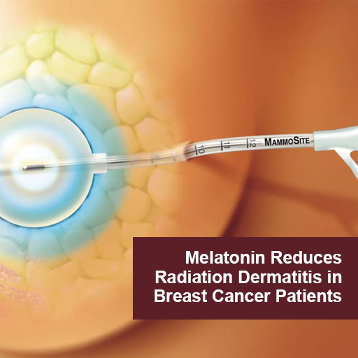
Melatonin Reduces Radiation Dermatitis in Breast Cancer Patients

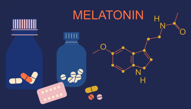
Radiation induced dermatitis is a common adverse effect and complication of radiotherapy.  Topical melatonin has proven to be a safe and effective agent in the prevention and treatment of radio-dermatitis in breast cancer patients.   Melatonin protects the skin by acting both as a topical anti-inflammatory and as an antioxidant. (5)

In a randomized placebo controlled trial women undergoing radiotherapy applied topical melatonin gel twice daily and for 2 weeks after completing radiotherapy.  The control group received a placebo gel. In the melatonin group  the occurrence of Grade1/2 radiation dermatitis was 59% vs 90% of the placebo group. “Patients treated with melatonin-containing emulsion experienced significantly reduced radiation dermatitis compared to patients receiving placebo.” (1). 

Topical Melatonin

  • Reduces inflammation and oxidative stress at the site of  irradiation
  • Delays onset of radio-dermatitis
  • Decreases the intensity of radiodermatitis and skin damage
  • Promotes resolution of dermatitis 
  • Promotes healing of skin
  • Improves Quality of Life 

Stress, nervous strain.Sleepless problem.Medicine diffuser.Flat vector

In two double blind randomized placebo controlled trials by the same research team, breast cancer patients applied a low dose melatonin cream twice daily during their course of radiotherapy.  The control group received a placebo cream.  During the first two weeks of therapy there was no difference between the 2 groups. Although there was no statistical difference, the researchers state that melatonin “demonstrated a protective effect”.   They posited that another study with a higher dose of melatonin is warranted. (3)  Researchers reported that melatonin decreased  breast symptoms of radiation dermatitis and improved quality of life for patients in the melatonin group (4)

In a case report a breast cancer patient applied melatonin cream daily and for 3 weeks after completing. radiotherapy treatment.  Radiation dermatitis did not appear at the radiated site until one week after final treatment (a delayed response) and resolved within 3 weeks of continued use of topical melatonin. (2)

There have been no reports of adverse effects with the use of  topical melatonin.

Apply topical melatonin to irradiated skin twice daily during radiotherapy and continuing for 2-3 weeks after completion of course of treatment until the skin is healed.

Melatonin gel or cream can be made by a compounding pharmacist.  Melatonin is also commercially available as a topical cream, as a liposomal solution and as a topical spray. 

References

1 Melatonin for Prevention of Breast Radiation Dermatitis: A Phase II, Prospective, Double-Blind Randomized Trial.

Ben-David MA, Elkayam R, Gelernter I, Pfeffer RM.

Isr Med Assoc J. 2016 Mar-Apr;18(3-4):188-92.

PMID: 27228641 Free article. Clinical Trial. 

2 Compounded Melatonin Cream for the Prevention and Treatment of Radiation Dermatitis: A Case Report.

Garcia-Segura LC, Garcia-Segura JC, Delgado DC, Romero MN, Salgado EC, Llorens LP.

Int J Pharm Compd. 2022 Jan-Feb;26(1):6-8.

PMID: 35081038 

3 Effect of melatonin cream on acute radiation dermatitis in patients with primary breast cancer: A double-blind, randomized, placebo-controlled trial.

Zetner D, Kamby C, Christophersen C, Gülen S, Paulsen CB, Piga E, Hoffmeyer B, Mahmood F, Rosenberg J.

J Pineal Res. 2023 Apr 13:e12873. doi: 10.1111/jpi.12873. Online ahead of print.

PMID: 37055944 


blood-test

What Common Blood Tests Can Detect Early Signs of Cancer

While there is no simple blood test for predicting who will get cancer, there is a lot of information to be gleaned from basic blood work that, taken together, reveals much about an individual’s predispositions for many forms of cancer. By monitoring selected biomarkers routinely measured in primary care, you can learn a lot about physiological patterns that promote carcinogenesis, proliferation, progression, and recurrence long before tumor markers emerge or there are radiological or pathological findings indicating cancer.

The art of assessment lies in part in recognizing the patterns. By learning how to read the multiple biochemical signals that emerge from a pro-carcinogenic “tumor microenvironment,” you can begin to practice real prevention, and give your patients the opportunity for significant improvements in both health-span and lifespan.

The tests included in this article here are ones you are routinely ordering in the integrative and functional medicine setting. While they are not to be misconstrued as diagnostic tests for cancer, they can indicate that a patient is at increased risk, and that further assessment and action is required to identify potential malignancy.

In people who’ve had cancer, these common tests are often prognostic for disease progression and recurrence.

It is vital that primary care practitioners do a better job of recognizing the early signs of recurrence among cancer survivors. According to the American Cancer Society’s 2016-2017 Survivorship Facts and Figures, the population of cancer survivors will increase to 20.3 million by January 1, 2026.

After conventional oncology treatment is finished, these patients typically return to their primary care physicians. They are highly motivated, ripe for change, and in search of clinicians who can support their efforts to restore health and prevent recurrence.

The tests described below will help you fill that role.

Complete Blood Count
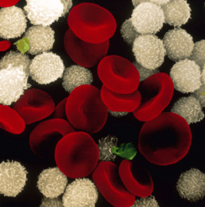
One of the most common biomarkers of overall health is the Complete Blood Cell panel, which can be used to monitor hematologic abnormalities caused by solid tumors, hematologic malignancies, as well as the side-effects of the therapies used to treat them.

The following findings are not definitive diagnostic signals, but taken together, they suggest that someone is at greatly increased risk:

  • Elevated White Blood Cells > 11.0 109/L
  • Elevated Platelets > 350 109/L
  • Low Hemoglobin <10.0 g/dL
  • High Neutrophil to Lymphocyte Ratio (NLR)

The latter finding—a high NLR—is especially important.

Neutrophils promote cancer progression, proliferation, and metastasis by increasing vascular endothelial growth factor (VEGF), Hepatocyte growth factors, inflammatory cytokines IL-6, IL-8, matrix metalloproteinases (MMP), and elastase. Neutrophils and macrophages secrete tumor growth promoting factors and contribute to a proliferative tumor microenvironment.

Therefore a high neutrophil count is suggestive of a neoplastic process somewhere in the body.

According to a 2014 metanalysis of 57 studies, an NLR greater than 4.0 was associated with a hazard ratio for overall survival (OS) of 1.81 (95% CI = 1.67 to 1.97; P < .001), an effect observed in all disease subgroups, sites, and stages and that predicts increased risk of mortality (Templeton AJ, et al. JNCI. 2014:106(6).)

Simply put, an NLR over 4 predicts tumor progression and poor overall survival. This is a readily available and inexpensive biomarker with a lot of prognostic value.

Hyperglycemia

A fasting glucose in the range of 100-126 mg/dl is suggestive of cancer risk.

Glucose may have a direct role in cancer development. Tumor cells have increased numbers of receptors for insulin, insulin-like growth factor, and GLUT4. Thus, they transport more glucose into themselves, and this promotes growth and proliferation. It is the main reason for using a low-glycemic, modified ketogenic diet in patients with cancer.

Proliferating tumor cells have up- regulated glucose transporters. Elevated serum glucose is linked to increased risk and progression of many solid cancers, including breast cancer (Haseen SD, et al. Asian Pac J Cancer Prev, 2015:  16, 675-8).

High glucose levels also result in a state of chronic inflammation, which leads to an increase of cytokines, such as interleukin 6 (IL-6), tissue necrosis factor alpha (TNF-α) and vascular endothelial growth factor (VEGF). All of these promote cancer progression, proliferation, and metastasis (Crawley DJ, et al. BMC Cancer, 14(1), 985).

Given the high prevalence of diabetes, metabolic syndrome, and insulin resistance in the US, this is an important indicator to watch.

Serum glucose is a modifiable risk factor. Diet and lifestyle changes that reduce and regulate glucose will also help to reduce risk and progression of cancer.

High Insulin & Low SHBG 

Prolonged hyperinsulinemia leads to reduced hepatic production of sex hormone binding globulin (SHBG). This, in turn, increases risk of steroid hormone driven cancers. Low SHBG results in increased amounts of unbound estrogens and androgens that drive carcinogenesis in breast, endometrial, prostate lung, colorectal and pancreatic tissues.

Free unbound estrogen also exerts immunosuppressive effects in the tumor microenvironment, and has a profound impact on anti-tumor immunity and tumor-promoting inflammation that is completely independent from its direct activity on tumor cells (Svoronos N, et al. Cancer Discovery, 2017: 7(1), 72-85).

Low Serum Albumin
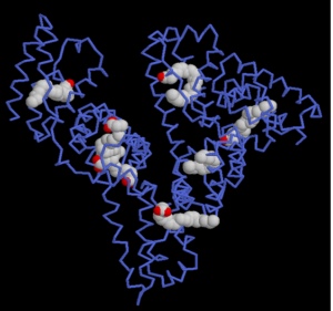
Serum albumin levels have prognostic significance in cancer, and can be used to better define baseline risk in cancer patients. It is generally useful in assessing the nutritional status, disease severity, disease progression, and prognosis.

In a multivariate analysis of 29 studies, Gupta and Lis found, “higher serum albumin levels to be associated with better survival.” (Gupta D, Lis CG. Nutrition Journal, 2010: 9(1), 69).

In the early stages of cancer, there is slight or no hypoalbuminemia. But as the disease progresses, malnutrition and inflammation suppress albumin synthesis, and albumin levels drop significantly.

Albumin levels under 3.5 g/dL are often seen in patients with sarcopenia and cachexia. Malnutrition is a predictor of reduced survival. It is also associated with deteriorating quality of life, decreased response to treatment, increased risk of chemotherapy-induced toxicity, and a reduction in cancer survival.

On the high side, albumin concentrations above 37.5 g/L are predictive of both chemotoxicity and of survival (Srdic D, et al. Supportive Care in Cancer, 2016: 24(11), 4495-4502).

It is also important to look at the Albumin-to-Globulin Ratio.

A ratio of less than 1.66 is a risk factor for cancer incidence and mortality, both short- and long term, in generally healthy screened adults. In people who’ve already developed some form of cancer, a low albumin-to-globulin ratio predicts low overall survival (Suh B, et al. Ann Ocol (2014): 25(11), 2260-2266).

Elevated Ferritin
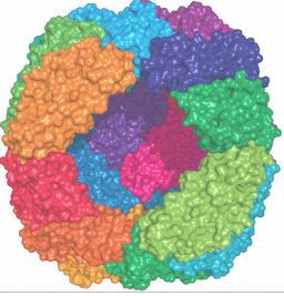
Ferritin, a strong negative survival predictor, has been associated with the pathological processes of inflammation and infection. High ferritin is suggestive of inflammation, immunosuppression, tumor angiogenesis, and proliferation.

Elevated serum ferritin—indicated by levels over 200 ng/ml in men, and over 150 ng/ml in women–have been seen in people with breast cancer, pancreatic cancer, non-small cell lung cancer, hepatocellular carcinoma, leukemia, colorectal cancer and lymphoma.

High ferritin levels are significantly associated with reduced survival time and increased mortality in cancer patients (Lee S, et al. J Cancer, 2016: 7(8), 957-964)

25-OH Vitamin D Deficiency

Vitamin D has a multi-functional impact on the tumor microenvironment. Increased levels of Vitamin D are associated with reduced occurrence and reduced mortality of different types of cancer, including skin, prostate, breast, colon, ovary, kidney, and bladder.

Vitamin D is involved in a very wide range of physiological processes relevant to cancer development, including: Regulation of Gene Transcription; Growth Arrest; Apoptosis; Cellular Differentiation; DNA Repair; Antioxidant Protection; Immune Modulation; Regulation of Pro-Inflammatory Cytokines; and Control of Angiogenesis & Metastasis.

Low or suboptimal levels of 25-OH Vitamin D are associated not only with increased risk of various forms of cancer, but also with poor prognosis, and more aggressive disease (McDonnell SL, et al. PloS One, 2016: 11(4), e0152441).

This is particularly true in breast cancer. In one study, vitamin D-deficient women with breast cancer typically had more aggressive molecular phenotypes and worse prognostic indicators than those with adequate vitamin D (Williams JD, et al. Endocrinology, 2016: 157(4), 1341-1347).

The Vitamin D Council suggests repletion to 40 to 80 ng/mL, with a target of 50 ng/ml, for optimal health on multiple fronts, including colorectal cancer prevention (Bischoff-Ferrari HA, et al. Am J Clin Nutr, 2006: 84(1), 18-28).

Supplementation to reach mean serum concentrations of 72 nmol/L showed a beneficial effect  against cancer development (Lappe JM, et al. Am J Clin Nutr. 2007: 85(6), 1586-1591).

When assessing patients in the context of cancer risk, the following guidelines are useful:

25 –hydroxy- Vitamin D (ng/ml)

Deficient                                                        < 50

Optimal                                                           50-70

Optimal for Cancer & CVD                70-99

Excess                                                            >100

Elevated Lactic Acid Dehydrogenase

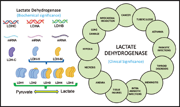
Lactate dehydrogenase (LDH) is an enzyme that catalyzes the reduction of pyruvate to lactate.

Aberrant metabolism and inefficient fuel production is a characteristic of tumor cells, which are dominated by aerobic glycolysis, increased lactate production, and a higher uptake of glucose (the Warburg effect).

Elevated LDH may be a marker of these aberrant metabolic processes in cancer cells.

The normal range for LDH is thought to be 100-333 u/L, with levels greater than 245 u/L considered to be in the upper quartile of normal. Above that 245 u/L mark, it is suggestive of early carcinogenesis, tumor cell proliferation, tumor progression, and poor prognosis.

It is often highly elevated in aggressive forms of cancer and hematological malignancies including: melanoma, lymphoma, acute leukemia, seminoma germ cell, pancreatic, gastric, lung, renal cell, nasopharyngeal, esophageal, cervical, and prostate cancers (Wulaningsih W, et al. Br J Cancer. 2015:113(9). Zhang J, et al. Sci Rep. 2015:5, 9800).

Elevated C-Reactive Protein
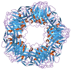
C-Reactive Protein (CRP) is a well-established inflammatory marker. It is also a biomarker of cancer survival.

CRP is elevated in patients with solid tumors, and high levels predict poor prognosis, blunted treatment response, as well as tumor recurrence.

As part of the systemic inflammatory response to a tumor, the body releases pro-inflammatory cytokines and growth factors. Interleukin-6, produced by the tumor or surrounding cells, stimulates liver production of acute-phase reaction proteins that increase C-reactive protein (CRP) and fibrinogen.

Elevated CRP correlates with disease stage and increased cancer mortality (Shrotriya S, et al. PloS One. 2015: 10(12), e0143080). Individuals with a high baseline CRP (>3 mg/L) have an 80% greater risk of early death compared with those with low CRP levels (<1 mg/L).  

Patients with invasive breast cancer and CRP levels>3 mg/L at diagnosis have a 1.7 fold increased risk of death compared to those with CRP levels<1 mg/L at diagnosis (Allin KH, et al. Breast Cancer Res. 2011: 13(3), R55).

Converging Signals

No one of the aforementioned test parameters is, in and of itself, an indicator that someone has cancer. But by looking at standard blood test results in a new way, you can start to recognize the patterns of high risk and active cancer physiology. This is crucial to early identification and early intervention.

Clinicians who are aware of the converging signs can meaningfully shift the microenvironment from one that promotes cancer to one that is not supportive of carcinogenesis, proliferation, or progression. In the same way, we can provide meaningful support for the rising tide of underserved cancer survivors and at-risk patients in need of not only a disease plan, but also a health plan.

Biomarker Lactic Acid Dehydrogenase Predicts Cancer Progression and Overall Survival

Aberrant metabolism and inefficient fuel production are characteristic of tumor cells, which are dominated by aerobic glycolysis, increased lactate production, and a higher uptake of glucose (the Warburg effect). Elevated LDH is a marker of these aberrant metabolic processes in cancer cells. High serum LDH levels are associated with poor prognosis in patients with cancer and predict progression and overall survival.

Aerobic glycolysis was described for the first time about a century ago by Otto H. Warburg who showed that cancer cells metabolize glucose differently than normal cells (Warburg effect) and that tumors derive energy mainly from the conversion of glucose to lactic acid and minimally via cellular respiration involving oxygen. Tumors produce massive amounts of the aerobic glycolysis waste product, lactic acid. This is evidence of deregulated metabolism, hence the understanding of cancer as “disorder of cellular metabolism”.  Lactic Acid itself may promote the growth and spread of cancer cells, especially at high concentrations by changing the tumor microenvironment.

Lactate dehydrogenase (LDH) is an enzyme that catalyzes the reduction of pyruvate to lactate at the end of the glycolytic pathway.

The normal range for LDH is 100-333 u/L, with levels greater than 245 u/L considered to be in the upper quartile of normal.  Elevated LDH, above 245 u/L, is suggestive of early carcinogenesis, tumor cell proliferation, tumor progression, and poor prognosis.

LDH is often highly elevated in aggressive forms of cancer and hematological malignancies including melanoma, lymphoma, acute leukemia, seminoma germ cell, pancreatic, gastric, lung, renal cell, nasopharyngeal, esophageal, cervical, and prostate cancers.

The OutSmart Cancer System® recognizes cancer as a metabolic syndrome and leverages the abnormal metabolism of tumor cells to exert influence over the tumor microenvironment and the behavior of tumor cells. Attending to the Cancer Terrain is a fundamental approach for influencing cancer cell metabolism.  

EGCG, a catechin found in Green Tea (H. Camellia sinensis) has been identified as an agent which inhibits LDH activity in normal and low oxygen environments by influencing the conversion of pyruvate to lactate at the end of the glycolytic pathway.  This may deprive cancer cells of their preferred fuel, glucose, and metabolites, including lactate that produces a favorable environment for malignant proliferation, growth, and progression. Recommended Therapeutic Dose 1-3 grams daily.

Monitoring trends in LDH is a method of both identifying abnormal cellular metabolism found in many solid and hematologic malignancies and is also of value in identifying early signs of recurrence as well as disease progression.

For patients achieving remission, during the first two years after completion of cancer treatment, LDH and other biomarkers of the Cancer Terrain are monitored every 3 months.  Thereafter, every six months for 3-10 more years to track and identify early signs of recurrence.  

For patients living with cancer as a chronic illness, LDH and biomarkers of the Cancer Terrain are monitored every 3 months to track evidence of recurrence and treatment resistance.


Learn more about monitoring the Cancer Terrain and the Tumor Microenvironment.
Receive training in Dr. Nalini’s OUTSMART CANCER SYSTEM ®.
www.aiiore.com

By using biomarkers of the Cancer Terrain and cellular metabolism, it is possible to identify trends that allow for early intervention. LDH is one of the most valuable and reliable biomarkers reflecting the active presence of the aberrant physiology of tumor cells and is prognostic and predictive of progression and overall survival in cancer patients.


Selected References:

Oral Cancer AIIORE Blog

Phytochemicals in Oral Cancer Prevention and Therapy

Oral cancers originate in the oral cavity and may spread to the neck and throat and local lymph nodes and can metastasize. These cancers are most commonly squamous cell carcinomas and are often very aggressive.

Cancers of the oral cavity, head, and neck are linked to drinking alcohol, smoking tobacco, betel nut chewing, human papillomavirus infection, and nutritional deficiencies.

Phytochemicals are a useful adjunct therapy for both prevention and therapy.

The continuous increase in cancer cases, the failure of conventional chemotherapies, and the excessive toxicity of chemotherapies demand alternative cancer treatments.

Phytochemicals can inhibit or antagonize factors, which are dysregulated in cancer cells and may enhance the effects of conventional therapy or could be developed into a stand-alone therapy*

Phytochemicals may exert their chemopreventive properties by blocking the critical events of tumor initiation and promotion, thereby reversing the premalignant stage. Phytochemicals may also prevent tumorigenesis by inhibiting or slowing tumor progression or promoting cell differentiation. Furthermore, phytochemicals can enhance innate immune surveillance and improve the elimination of transformed cells.”**

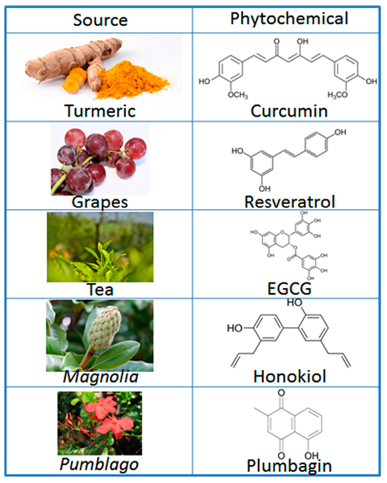
Phytochemicals that impact multiple pathways active in the development, growth, progression, and spread of oral cancers include

Phytochemicals in Oral Cancer
  • Black Raspberry anthocyanins
  • Green Tea Catechins (EGCG, EGC, ECG)
  • Curcuma longa (curcuminoids) (tumeric)
  • Alliums: Garlic and Onions (allicin, s-allylcysteine)
  • Resveratrol 3,4’,5-trihydroxy-trans-stilbene
  • Lycopene carotenoid (tomatoes, red bell peppers)
  • Astaxaxanthin and Canthaxanthin  carotenoid xanthophylls  (green leafy vegetables)
  • Bromelain cysteine protease (pineapple)

For a detailed and thorough discussion of risk factors, etiologies, signs and symptoms, histopathology, molecular mechanisms and therapeutic interventions in oral, head, and neck cancers see: 

*Tzu-Ying Lee , Yu-Hsin Tseng Review : Biomolecules  The Potential of Phytochemicals in Oral Cancer Prevention and Therapy: A Review of the Evidence 2020 Aug 6;10(8):1150. doi: 10.3390/biom10081150

** Kotecha R., Takami A., Espinoza J.L. Dietary phytochemicals and cancer chemoprevention: A review of the clinical evidence. Oncotarget. 2016;7:52517–52529. doi: 10.18632/oncotarget.9593.


Let The Oncologist Be The Disease Expert. Become The Health Expert That Cancer Patients Are Looking For.

You may not treat cancer in your practice, but you do have patients who are at risk due to personal and family history, patients who may be undergoing or recovering from treatments, patients who are survivors worried about recurrence and patients living with cancer as a chronic illness.  And you may also have patients who are family members concerned about their loved ones. 


There is no HEALTH MODEL in conventional oncology care, yet health and wellbeing, peace of mind and sense of agency are in the center of the hearts and minds of cancer patients, cancer survivors and their families. 


There will be 19 million cancer survivors in the US alone by 2024.  Who is supporting their health?  Who is trained to help them recover and keep them well??  …not the oncologist.


How can you help these patients?

A  breast cancer survivor who successfully completed her treatments 8 years ago comes into your office as a new patient complaining of persistent peripheral neuropathy and ongoing cognitive changes since her treatment.  How can you resolve these long-term adverse effects?


An ovarian cancer patient currently undergoing aggressive treatment every 21 days comes into your office complaining of severe diarrhea, neuropathy and sleep disruption.  What can you do to help her get through her treatments with less adverse effects, maintain her weight and nutritional status?


A colorectal cancer survivor who completed his treatment 3 months ago is continuing to have 10-15 bowel movements daily and is profoundly fatigued.  What will you do to restore normal bowel function?


A prostate cancer patient on endocrine blockade therapy is suffering from
hot flashes. Should you also be concerned about loss of bone mass and sleep cycle disruption?


An endometrial cancer survivor is suffering from dermatitis and colitis, adverse effects of her dramatically successful immunotherapy treatment and now has chronic autoimmune inflammation. How will you manage this?


A head and neck cancer patient who has trouble swallowing is losing weight and muscle mass.


How can you provide a plan for repair from oral mucositis, restoration of the oral mircrobiome and repletion of calories and nutrients?


These patients are searching for clinicians that can guide and support them through every phase of their cancer journey.  Just as in helping your patients navigate other chronic illnesses, patients look to you for a plan, for monitoring and guidance so that they can maintain and regain their health during and after their treatments.


When a patient has a collaborative team providing integrative care everyone wins, the patients, families and care providers.  Patients who have a clear plan and support have the opportunity for better outcomes, better prognosis, greater peace of mind, a sense of control and agency and an improved quality of life. 


Let the oncologist be the cancer expert. You can be the health expert on their team.


Standard of care in oncology must  change such that care includes not only a team of disease experts (usually medical oncologist, surgeon, radiologist) but ALSO a team of health experts.


Towards this end  I founded the American Institute of Integrative Oncology Research and Education and  have created an online self-paced training program for front line clinicians who want to expand their skills and their practice and  fill the huge need in our communities and serve these patients.  If you did not specialize in oncology, you probably had one course on this topic but you need to fill the gap in your training to feel confident in doing so.


The Foundations of Integrative Oncology Training is not for clinicians who want to practice oncology.  It is front-line clinicians who want to feel confident, knowledgeable and well trained in supporting the health side of the cancer equation. This self- paced online training is for clinicians who want to increase their impact, expand and grow their practice and represents 35 years of clinical practice and experience.


The first step is learning how to take a comprehensive and complete history of patients whose lives have been touched by cancer.  


You can receive a complimentary copy of the

OUTSMART CANCER CARE PLANNER History and Intake Form

and learn more about the Foundations of Integrative Oncology training here


OutSmart Cancer Care Planner