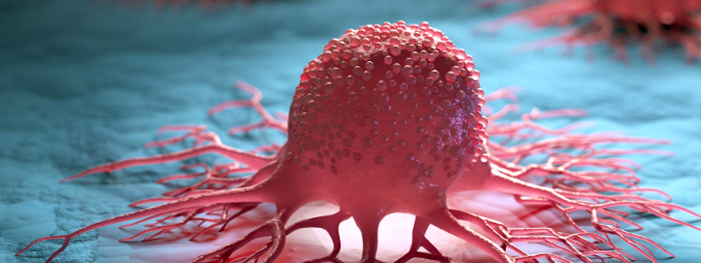
Home » Archives for Admin AIIORE » Page 4

Author: Admin AIIORE

green-tea-extract

Green Tea Extract Reduces Severity of Radiation Dermatitis

The use of a solution containing a green tea extract has been shown to reduce both the incidence and severity of radiation-induced dermatitis in women undergoing adjunctive radiotherapy for breast cancer. This was the conclusion of a phase 2 randomized, placebo-controlled trial by a team of Chinese researchers.

Data from the World Health Organization indicates that in 2020, there were 2.3 million women diagnosed with breast cancer. In the treatment of women with breast cancer, radiation therapy is widely used conjunction with other therapies such as surgery, chemotherapy and hormonal therapies.  A common and frequent adverse effect of radiotherapy is radiation-induced dermatitis (RID) suffered by millions of women.

green-tea

The purpose of the current  study was to investigate the safety, tolerability and preliminary effectiveness of topical epigallocatechin-3-gallate (EGCG) for radiation dermatitis in patients with breast cancer receiving adjuvant radiotherapy.

A  solution of green tea extract  sprayed on the radiated areas of the skin reduced severity of radiation-induced dermatitis.

The Chinese team recruited women with breast cancer undergoing postoperative radiotherapy and randomized them (2:1) to receive either the green tea extract or placebo (normal saline solution).  These solutions were sprayed to the whole of the radiation field from the first day of therapy until two weeks after completion of treatment. 

A total of 165 women with a median age of 46 years were enrolled and randomized to EGCG, the primary catechin found in green tea or placebo.

The onset of radio-dermatitis was delayed by 2-3 weeks and the intensity and severity of the symptoms were significantly decreased in the treated group.  No skin toxicity was observed.

The authors concluded that prophylactic use of a green tea extract significantly reduced both the incidence and severity of RID and that it has the potential to become a new choice for skin care in women receiving radiotherapy.

Topical green tea extract supports restoration of skin integrity and control of inflammatory cytokines and oxidative stress in the skin. Green tea extract also reduces the acute skin-induced reactions including pain and sensations of burning, itching, pulling and tenderness.

Dr. Chilkov: Practical Application:

green-tea-leavesTopical Green Tea Extract Spray

To make a medicinal water extract: Place 8 organic green tea bags into a 16 oz glass jar or glass container.  Pour boiling water over the tea bags, cover immediately and steep for one hour.   After it has cooled to room temperature store covered in the refrigerator.  When ready to use transfer water extract to a a glass spray bottle.  Apply liberally to the radiation field before and after each radiotherapy session and three times daily for 3 weeks after the last radiotherapy session.  

Fresh Aloe Vera Gel poultice

Areas where skin is most impacted can be covered with. mashed fresh aloe vera gel and covered with a large gauze bandage.   This can easily be held in place underneath a sports bra or leotard or similar.  Apply fresh aloe gel twice daily. Allow to be in contact with the skin for several hours or overnight.  If you do not have access to a live aloe vera plant or fresh aloe gel you can use alcohol free aloe vera juice or aloe vera gel commonly found in natural foods stores.    Aloe Vera is the botanical of choice for repair of radiation damaged skin.

Topical Calendula Oil (not extract) is also a soothing topical anti-inflammatory agent for radiation induced dermatitis.  If the skin is very damaged, saturate a 4x4” gauze square and place over the affected area.

References:

Zhao H et al. 

Efficacy of Epigallocatechin-3-Gallate (EGCG)in Preventing Dermatitis in Patients With Breast Cancer Receiving Postoperative Radiotherapy: A Double-Blind, Placebo-Controlled, Phase 2 Randomized Clinical Trial JAMA Dermatol 2022

Zhao H, et al. 

Phase I study of topical epigallocatechin-3-gallate (EGCG) in patients with breast cancer

receiving adjuvant radiotherapy. Br J Radiol 2016; 89: 20150665.

Kyle T. Amber, BS et al

The Use of Antioxidants in Radiotherapy-Induced Skin Toxicity 

Integrative Cancer Therapies 2014, Vol. 13(1) 38–45

Book Review: You Finished Treatment-Now What?


A Field Guide for Cancer Survivors.
By Dr. Amy Rothenberg

You-Finished-Treatment-Now What

You Finished Treatment, Now What? A Field Guide for Cancer Survivors by Dr. Amy Rothberg is a roadmap for lifestyle and natural medicine approaches to address health challenges that persist after cancer care, and to reduce the risk of recurrence.

Dr Rothenberg wrote this guide for cancer survivors and those on their support and care team.

You Finished Treatment, highlights the evidence for an integrative approach to healing that Dr. Rothenberg has used for over 37 years practicing as a licensed naturopathic doctor.

She is also a breast and ovarian cancer survivor/thriver herself. She wrote this book to make sense of an overwhelming topic, in a user-friendly, accessible way, providing both actionable information and inspiration.

As a survivor/thriver of both ovarian cancer and breast cancer she speaks both from the physician’s and the patient’s point of view with heart, levity and solid, practical advice.

When diagnosed with cancer in 2014, Dr. Rothenberg sought and received state-of-the-art care at a renowned teaching hospital and had her own naturopathic medical team to help her best handle treatment, and rebound afterward.

Her writing is evidence-informed, while also bringing her personal experience as a doctor, patient, wife, mother, sister, and friend. Offering a natural, integrative medicine perspective on items in the news, find Dr.

This is an example of the principles employed in Dr. Chilkov’s OutSmart Cancer® System which is an integrative approach to combining the best of modern oncology with the best of research informed modern and traditional naturopathic systems of medicine for the very best outcomes.

This approach allows patients to have not only a plan for their disease, but also a plan to support their health during and after treatment and to support recovery, restoration and rejuvenation in support of both healthspan and lifespan.

Dr. Rothenberg’s both personal experiences and medical expertise combine to form a heartful and pragmatic approach with clear guidelines and recommendations. This book is a wonderful resource for both patients and families as well as care providers.

Breast-Cancer

Changing the Management of Cancer with Personalized Testing


Personalized cDNA surveillance for patients with high-risk breast cancer

Is there a more sensitive technology that can detect preclinical breast cancer progression?

It is now possible to monitor fragments of cell free tumor DNA (ctDNA) circulating in the blood. This falls under the umbrella of “liquid biopsies” which monitor tumor burden, tumor response to treatment and early signs of recurrence or progression without a scan or need for a new surgical or biopsy tissue sample.

  • “Up to 30% of patients with breast cancer relapse after primary treatment.
  • There are no sensitive and reliable tests to monitor these patients and detect distant metastases before overt recurrence.
  • Breast cancer cell free tumor DNA blood test (liquid biopsy) can detect recurrence up to 2 years earlier than currently available conventional serum tumor markers and radiologic studies.
  • Cell free tumor DNA assays predict breast cancer recurrence earlier and with greater accuracy than traditional tools by using a highly-personalized molecular residual disease assay.

A cell free tumor DNA (ctDNA) assay is a personalized, tumor-informed assay with the power to give you earlier, clearer insight into your patient's disease. By detecting and quantifying ctDNA, you can optimize your ability to assess risk, predict recurrence, and monitor treatment response in those most at risk for progression.

This technology can be used to monitor a wide range of cancers. While this type of monitoring has not yet been widely adopted as “standard of care” I encourage you to educate all of your patients and their care providers to adopt the use of this highly reliable screening tool now.

Here, we demonstrate the use of personalized circulating tumor DNA (ctDNA) profiling for detection of recurrence in breast cancer.”

Cell free tumor DNA assays use a sample of the patient’s tumor tissue to develop a unique DNA fingerprint. After that, follow-up blood draws capture changes in the level of ctDNA, giving clinicians a better picture of a patient’s risk of recurrence without the need for another tissue sample and may decrease the need for frequent scans and repeated frequency of exposure to radiation and contrast material.

neodjuvant

A recent study “demonstrates that patient specific ctDNA analysis can be a sensitive and specific approach for disease surveillance for patients with breast cancer. More importantly, earlier detection of up to 2 years provides a possible window for therapeutic intervention. “(1)

Currently, there are no sensitive and specific clinical tests available to follow patients with breast cancer after primary treatment. Signatera developed a patient-specific method to analyze circulating tumor DNA (ctDNA) that allows for monitoring of these patients regardless of molecular genotype. In this study, we analyzed 208 blood samples from 49 patients monitored longitudinally for up to 4 years after completion of adjuvant chemotherapy to determine whether personalized ctDNA assays can allow for more effective monitoring than current clinical tests such as CA 15-3. Remarkably, for the patients that recurred, our test detected molecular relapse up to 2 years ahead of clinical relapse (median, 8.9 months) with 89% sensitivity and 100% specificity. This may provide a critical window of opportunity for additional therapeutic intervention.” (1)

hope

Data from a retrospective cohort analysis of EBLIS, a study designed to determine the lead interval between ctDNA detection and clinical metastatic disease, and to determine whether ctDNA in plasma can detect recurrent disease earlier than traditional methods, demonstrated that Signatera can accurately predicts metastatic relapse with a significant lead time over imaging and CA 15-3 (200 days on average)

Neoadjuvant

“…our study shows promise that early response prediction by highly sensitive ctDNA analysis in high-risk early breast cancer patients may facilitate a timely and judicious change in treatment to improve patients’ chances of achieving favorable long-term outcomes.(2)

Surveillance

Patients undergoing treatment as well as those who have completed their course of treatment can be assessed both for response to treatment during a course of therapy as well as for early signs of reurrence after treatment has been completed. In a study of patients undergoing treatment with Pembrolizumab, a checkpoint inhibitor.

“Baseline ctDNA concentration correlated with progression-free survival, overall survival, clinical response and clinical benefit. This association became stronger when considering ctDNA kinetics during treatment. All 12 patients with ctDNA clearance during treatment were alive with median 25 months follow up. This study demonstrates the potential for broad clinical utility of ctDNA-based surveillance in patients treated with ICB.” (3)

Recommended labs offering this technology include Natera, INVITAE, Foundation One, Caris Life Sciences. All of these labs are highly regarded in the oncology community. (Disclosure: I have no financial relationships with any of the labs recommended in this article.)

How often should these assays be performed?
I recommend monitoring monthly during active treatment to determine if the current treatment is effective and continuing to be effective. This is a way to identify treatment resistance early.

top10-badgeI recommend monitoring every three months during the first two years after completing treatment or for patients with advanced receiving ongoing treatments. (For example advanced breast cancer patients receiving hormonal treatments, immunotherapy treatments, checkpoint inhibitor treatments or chemotherapy treatments over long periods of time.)

For long term survivors I recommend monitoring every 6 months until the 10 year No Evidence of Disease anniversary.

This is the same schedule of monitoring that we use in the OUTSMART CANCER® System to follow measurable biomarkers in the tumor microenvironment.

Discover how you can join
Foundations of Integrative Oncology Professional Online Training
Master the OUTSMART CANCER® System
Receive Monthly Live Mentoring and Case Supervision
from Dr. Nalini Chilkov
CLICK HERE

References

  1. Coombes C, Page K, Salari R, et al. Personalized Detection of Circulating Tumor DNA Antedates Breast Cancer Metastatic Recurrence. Clinical Cancer Research. 2019;25(14):4255-4263.
  2. Circulating tumor DNA in neoadjuvant-treated breast cancer reflects response and survival M. J. M. Magbanua. https://doi.org/10.1016/j.annonc.2020.11.007
  3. Bratman, S.V., Yang, S.Y.C., Iafolla, M.A.J. et al. Personalized circulating tumor DNA analysis as a predictive biomarker in solid tumor patients treated with pembrolizumab. Nat Cancer 1, 873–881 (2020). https://doi.org/10.1038/s43018-020-0096-5
  4. https://www.natera.com/info/know-breast-cancer/?utm_source=cancer-therapy-advisor&utm_medium=email&utm_campaign=breast-cancer-launch
Probiotics

Oral Probiotics Reduce Complications of Surgery


Using probiotics before surgery prepares the patient for post operative stressors and complications. Using probiotics after surgery continues the support for the microbiome post operatively.

It is my practice to administer oral probiotics both before and after surgery with all of my patients.

Overall, using probiotics as part of pre-op and post-op care offers the following benefits

  • Reduction in Pro-Inflammatory Cytokines
  • Prevention of Surgical Infection and Sepsis 
  • Promotion of gastrointestinal microbial balance
  • Amelioration of adverse effects of oral antibiotics 
  • Decrease in adverse effects of opioids on gastrointestinal function
  • Promotion of Wound Healing at the surgical site

Use of oral probiotics is well tolerated and safe for use not only in cancer related surgeries but in a wide range of surgical procedures. 

Researchers conducting a randomized double blind placebo controlled study on the post operative effects of oral probiotics in patients undergoing resection for colorectal cancer concluded that probiotics not only decrease rates of infection at the incision site, respiratory and urinary systems but also inhibit proinflammatory factors such as TNFa, IL-17A , IL-17C, IL-22, IL-10 and IL-12.   Subjects in the treatment arm were given a 30 billion CFU mixture of six viable strains of Lactobacillus acidophilus, L. lactis, L. casei, Bifidobacterium longum, B. bifidum, and B. infantis twice daily for 6 months beginning 4 weeks postoperatively. [NB: I recommend starting pre-operatively].   Subjects in this arm did not experience infection, diarrhea or require antibiotics.

Zaharuddin L, Mokhtar NM, Muhammad Nawawi KN, Raja Ali RA. A randomized double-blind placebo-controlled trial of probiotics in post-surgical colorectal cancer. BMC Gastroenterol. 2019 Jul 24;19(1):131. doi: 10.1186/s12876-019-1047-4. PMID: 31340751; PMCID: PMC6657028.

In another study of patients receiving abdominal surgeries  oral probiotics were administered for 8 weeks.  The strains included were  L. plantarum, L. lactis, and L. delbrueckii. The study found statistically significant postoperative treatment reductions in abdominal pain and bloating, and significant improvements in stool formation. No clinically relevant adverse events were reported, and the treatment was well-tolerated by all patients. 

Bonavina L, Arini A, Ficano L, Iannuzziello D, Pasquale L, Aragona SE, Ciprandi G, On Digestive Disorders ISG. Post-surgical intestinal dysbiosis: use of an innovative mixture (Lactobacillus plantarum LP01, Lactobacillus lactis subspecies cremoris LLC02, Lactobacillus delbrueckii LDD01). Acta Biomed. 2019 Jul 10;90(7-S):18-23. doi: 10.23750/abm.v90i7-S.8651. PMID: 31292422; PMCID: PMC6776165.

In a recent 2021 Review of 14 studies of patients receiving gastrointestinal surgeries, a disruption of intestinal microbiome is identified and the prevalence of specific bacteria had significantly changed after surgery.

Ferrie S, Webster A, Wu B, Tan C, Carey S. Gastrointestinal surgery and the gut microbiome: a systematic literature review. Eur J Clin Nutr. 2021 Jan;75(1):12-25. doi: 10.1038/s41430-020-0681-9. Epub 2020 Jul 13. PMID: 32661352.

Another Review of 10 studies also identified post operative changes in the composition of the intestinal microbiome in patients receiving gastrointestinal surgeries  and posits that complications after gastrointestinal surgeries are linked to changes in the composition of the gut flora.

Lederer, A. K., Pisarski, P., Kousoulas, L., Fichtner-Feigl, S., Hess, C., & Huber, R. (2017). Postoperative changes of the microbiome: are surgical complications related to the gut flora? A systematic review. BMC surgery, 17(1), 125. https://doi.org/10.1186/s12893-017-0325-8

A study on the use of specific probiotics in patients undergoing resection for  colorectal cancer concluded that inflammatory cytokines and serum zonulin levels significantly decreased with probiotics. Probiotic ingestion resulted in compositional changes in gut microbiota; greater increases and decreases in healthy vs pathogenic bacteria, respectively, occurred with probiotics. Compositional increase in healthy bacteria was associated with reduced white blood cells, neutrophils, neutrophil-lymphocyte ratio, and zonulin. Bifidobacterium composition was negatively correlated with zonulin levels in the probiotic group, indicating repair of intestinal epithelium as an effective barrier. Probiotics improved postoperative flatus control and modified postoperative changes in microbiota and inflammatory markers.   In this study oral probiotics were administered both pre-op and post-op.  Probiotic supplementation included a mixture of three probiotic strains (Bifidobacterium animalis subsp. lactis HY8002 (1 × 10⁸ cfu), Lactobacillus casei HY2782 (5 × 10⁷ cfu), and Lactobacillus plantarum HY7712 (5 × 10⁷ cfu)

Park, I. J., Lee, J. H., Kye, B. H., Oh, H. K., Cho, Y. B., Kim, Y. T., Kim, J. Y., Sung, N. Y., Kang, S. B., Seo, J. M., Sim, J. H., Lee, J. L., & Lee, I. K. (2020). Effects of PrObiotics on the Symptoms and Surgical ouTComes after Anterior REsection of Colon Cancer (POSTCARE): A Randomized, Double-Blind, Placebo-Controlled Trial. Journal of clinical medicine, 9(7), 2181. https://doi.org/10.3390/jcm9072181

immunotherapy

Preventing Abdominal Radiation Enteritis and Promoting Quality of Life in Gynecological Cancer Patients


Insulin and fructo-oligosaccharide prevent acute radiation enteritis in patients with gynecological cancer and improve quality-of-life

Insulin

Background/objectives: The pathogenesis of enteritis after abdominal radiotherapy (RT) is unknown, although changes in fecal microbiota may be involved. Prebiotics stimulate the proliferation of Lactobacillus spp and Bifidobacterium spp, and this may have positive effects on the intestinal mucosa during abdominal RT.

Subjects/methods: We performed a randomized, double-blind, placebo-controlled trial involving patients with gynecological cancer who received abdominal RT after surgery. Patients were randomized to receive prebiotics or placebo. The prebiotic group received a mixture of fiber (50 inulin and 50% fructo-oligosaccharide), and the placebo group received 6 g of maltodextrin twice daily from 1 week before to 3 weeks after RT. The number of bowel movements and stool consistency was recorded daily. Diarrhea was evaluated according to the Common Toxicity Criteria of the National Cancer Institute. Stool consistency was assessed using the 7-point Bristol scale. Patients' quality-of-life was evaluated at baseline and at completion of RT using the EORTC-QLQ-C30 (European Organization for Research and Treatment of Cancer quality-of-life Questionnaire C30) test.

Results: Thirty-eight women with a mean age of 60.3±11.8 years participated in the study. Both groups (prebiotic (n=20) and placebo (n=18)) were comparable in their baseline characteristics. The number of bowel movements per month increased in both groups during RT. The number of bowel movements per day increased in both groups. The number of days with watery stool (Bristol score 7) was lower in the prebiotic group (3.3±4.4 to 2.2±1.6) than in the placebo group (P=0.08). With respect to quality-of-life, the symptoms with the highest score in the placebo group were insomnia at baseline and diarrhea toward the end of the treatment.

bowel movements

In the prebiotic group, insomnia was the symptom with the highest score at both assessments, although the differences were not statistically significant.

Conclusions: Prebiotics can improve the consistency of stools in gynecologic cancer patients on RT. This finding could have important implications in the quality-of-life of these patients during treatment.

Vitamin-D

Higher Vitamin D Intake Reduces Risk of Colorectal Cancer

In a recent (2021) study* investigators concluded that higher total vitamin D intake is associated with decreased risks of
young-onset colorectal cancer and precursors (polyps).

colorectal-cancer
Colorectal Cancer (CRC) infographic for education
Colorectal Cancer (CRC) infographic for education illustration


Excluding skin cancers, colorectal cancer is the third most common cancer diagnosed in both men and women in the United States. The rate of people being diagnosed with colon or rectal cancer each year has dropped overall since the mid-1980s, mainly because more people are getting
screened (colonoscopy) and changing their lifestyle-related risk factors (healthy BMI, decreasing red meats, refined foods, and increasing fiber and phytochemicals from fruits and vegetables and whole grains).

From 2013 to 2017, incidence rates dropped by about 1% each year. But this downward trend is mostly in older adults and masks rising incidence among younger adults since at least the mid-1990s. From 2012 through 2016, it increased every year by 2% in people younger than 50 and 1% in people 50 to 64. 
https://www.cancer.org/cancer/colon-rectal-cancer/about/key-statistics.html

During the period from 1991 to 2015 the researchers* documented 111 cases of young-onset colorectal cancer and 3,317 colorectal polyps. Analysis showed that higher total vitamin D intake was associated with a significantly reduced risk of early-onset colorectal cancer. The same link was found between higher vitamin D intake and risk of colon polyps detected before age 50.

According to principal researcher K Ng, “Our results further support that vitamin D may be important in younger adults for health and possibly colorectal cancer prevention,

Understanding risk factors that are associated with young-onset colorectal cancer leads to informed recommendations about diet and lifestyle, as well as identifying high-risk individuals to target for earlier screening.

Many cancers, including colorectal and ovarian cancers, that were historically prevalent in older age groups are increasingly being seen in younger patients. Therefore, frontline, primary care providers, particularly in a health-focused setting such as functional, integrative, naturopathic, nutritional, and oriental medicine clinics MUST include patient teaching and appropriate screening in patients under 50.

Vitamin D is both a prognostic and predictive biomarker for both well patients and patients with a diagnosis or history of cancer. It is an important modulator of immunity and cancer biology in multiple histological types of cancer including skin, prostate, breast, ovary, colon, bladder, and kidney malignancies.

With regard to the functions of Vitamin D in the tumor microenvironment, Vitamin D

  • Regulates Gene Transcription
  • Induces Growth Arrest
  • Induces Apoptosis
  • Enhances DNA Repair
  • Enhances Antioxidant Protection
  • Enhances Immune Modulation
  • Enhances Differentiation
  • Decreases Pro-Inflammatory Cytokines
  • Decreases Invasion into the Extracellular matrix
  • Decreases Angiogenesis & Metastasis

I recommend including Serum 25-OH Vitamin D assays for ALL patients. Aim for Optimized Serum 25-OH Vitamin D levels of 60-80 ng/ml for promoting a robust cancer terrain that is inhospitable to the development, progression, and spread of cancer. Oral Vitamin D should be administered as Vitamin D3 cholecalciferol (not ergocalciferol, Vitamin D2).

*Reference: Hanseul Kim, Marla Lipsyc-Sharf, Xiaoyu Zong, Xiaoyan Wang, Jinhee Hur, Mingyang Song, Molin Wang, Stephanie A. Smith-Warner, Charles Fuchs, Shuji Ogino, Kana Wu, Andrew T. Chan, Yin Cao, Kimmie Ng, Edward L. Giovannucci.Total Vitamin D Intake and Risks of Early-Onset Colorectal Cancer and Precursors. Gastroenterology, 2021; DOI: 10.1053/j.gastro.2021.07.002

Download Dr. Chilkov's History and Intake Form for Cancer Patients and Survivors!

OutSmart Cancer


  • Confidently obtain a detailed cancer history
  • Address the unique needs of your patients whose lives have been touched by cancer
  • Create care plans focused on the post-treatment concerns of cancer survivors
  • Become the long-term health partner that patients in your community are seeking 
Cheating-cancer-Athena-Aktipis

How Evolution Helps Us Understand and Treat Cancer

The Cheating CellThe Cheating Cell: How Evolution Helps Us Understand and Treat Cancer - by Athena Aktipis

A fundamental and groundbreaking reassessment of how we view and manage cancer

When we think of the forces driving cancer, we don’t necessarily think of evolution. But evolution and cancer are closely linked, for the historical processes that created life also created cancer. The Cheating Cell delves into this extraordinary relationship, and shows that by understanding cancer’s evolutionary origins, researchers can come up with more effective, revolutionary treatments.


Athena Aktipis goes back billions of years to explore when unicellular forms became multicellular organisms. Within these bodies of cooperating cells, cheating ones arose, overusing resources and replicating out of control, giving rise to cancer.

Aktipis illustrates how evolution has paved the way for cancer’s ubiquity, and why it will exist as long as multicellular life does.

Even so, she argues, this doesn’t mean we should give up on treating cancer—in fact, evolutionary approaches offer new and promising options for the disease’s prevention and treatments that aim at long-term management rather than simple eradication.

Looking across species—from sponges and cacti to dogs and elephants—we are discovering new mechanisms of tumor suppression and the many ways that multicellular life-forms have evolved to keep cancer under control.

By accepting that cancer is a part of our biological past, present, and future—and that we cannot win a war against evolution—treatments can become smarter, more strategic, and more humane.

Unifying the latest research from biology, ecology, medicine, and social science, The Cheating Cell challenges us to rethink cancer’s fundamental nature and our relationship to it.

Grab Your Copy Here

blood-test

Selected Prognostic Cancer Biomarkers from Common Blood Tests

What Can You Learn About Cancer Survival from a CBC?

Common Biomarkers, Cancer Progression and Survival

tumor microenvironment

An excellent paper "Inflammatory markers in cancer: Potential resources * is a thorough and detailed discussion of routinely measured Cancer Biomarkers found in the inflamed tumor microenvironment that are indicative of immune capacity and prognosis. Clinicians will typically have access to a current CBC (Complete Blood Count) with differential and can readily calculate the lymphocyte ratios below

The presence of inflammatory markers is linked to both risks of cancer development and cancer survival.  Cancer-related inflammation is associated with tumorigenesis and tumor progression. Increased levels of multiple biomarkers are present in the tumor microenvironment.

A thorough evaluation will include Cytokines, Leukocytes, Acute-phase proteins (ferritin, ceruloplasmin, CRP).     

Although not routinely measured, prostaglandins, cyclooxygenases, lipoxygenases, transcription factors, and LDH may be elevated. 

Cancer Related Inflammation Promotes

  • Tumor Growth
  • Proliferation
  • Progression
  • Angiogenesis  
  • Metastasis
  • Thrombus Formation
  • Immune Suppression
  • Cancer Related Fatigue  
  • Depression
  • Pain

Selected Prognostic Cancer Biomarkers from the CBC and CMP

  • Neutrophil: Lymphocyte Ratio
  • Lymphocyte: Monocyte Ratio
  • Platelet: Lymphocyte Ratio
  • CRP: Albumin Ratio
  • Hypoalbuminemia

Neutrophil / Lymphocyte Ratio (NLR)

  • Neutrophyl LymphocytesThe neutrophils act as tumor-promoting leukocytes, capable of suppressing anti-tumor immune response; are effectors of angiogenesis; promote leakage of tumor cells and endothelial cells into the circulation, therefore contributing to participate in the metastatic cascade. Therefore, an elevated neutrophil count can stimulate tumor angiogenesis and contribute to disease progression, thus leading to a negative correlation between neutrophil density and patient survival. 
  • On the other hand, lymphocytes are a part of the host’s antitumor response the presence of lymphocytes in the tumor is associated with better responses to chemotherapy and better prognosis. 
  • Thus, the NLR can reflect the balance between the activation of the inflammatory pathway and the antitumor immune function. 
  • The division of neutrophil count by lymphocyte count is defined as NLR. 
  • An increase in NLR has been reported to correlate with poor prognosis in cancer patients.
  • A cut-off value between 2-4 has been reported in cancer patients.

Faria, S. S., Fernandes, P. C., Silva, M. J., Lima, V. C., Fontes, W., Freitas-Junior, R., Eterovic, A. K., Forget, P. The neutrophil-to-lymphocyte ratio: a narrative review. Ecancermedicalscience, 10, 702 (2016) DOI:10.3332/ecancer.2016.702 

Lymphocyte/Monocyte Ratio (LMR) 

  • Lymphocytopenia has been associated with increased tumor burden and poor prognosis. The probable cause could be the destruction of lymphocytes by tumor cells which decreases the body’s anti-tumor response.
  • Monocytosis has also been found to be associated with poor prognosis as they Tumor-associated macrophages, which are an important mediator of cancer progression and metastases. 
  • The division of lymphocyte count by monocyte count is defined as LMR. 
  • A low LMR as a simple biomarker of the host immune system has been suggested to be related to poor prognosis in various cancers. 
  • The median cut-off value for LMR has been reported to be 3.0.

Nishijima TF, Muss HB, Shachar SS, Tamura K, Takamatsu Y Prognostic value of the lymphocyte-to-monocyte ratio in patients with solid tumors: a systematic review and meta-analysis. Cancer Treat Rev 41(10) 971-8 (2015) DOI: 10.1016/j.ctrv.2015.10.003 

Platelet/Lymphocyte Ratio (PLR)

  • Platelet/ LymphocytePlatelets are another important tumor-promoting leukocyte. They secrete vascular endothelial growth factor (VEGF), epidermal growth factor (EGF), platelet-derived growth factor (PDGF), hepatocyte growth factor (HGF), insulin-like growth factor (IGF), transforming growth factor β (TGFβ), and many cytokines which promote epithelial to mesenchymal transition (EMT) and promote metastasis.
  • Lymphocytes, as we know, are part of the host's defense against tumors. 
  • The division of platelet count by lymphocyte count is defined as PLR. 
  • The cutoff value estimated for PLR is 160. 
  • A high PLR value correlates with a poor response to therapy and a bad prognosis.

Templeton AJ, Ace O, McNamara MG, Al-Mubarak M, Vera-Badillo FE, Hermanns T, Seruga B, Ocaña A, Tannock IF, Amir E. Prognostic role of platelet to lymphocyte ratio in solid tumors: a systematic review and meta-analysis. Cancer Epidemiol Biomarkers Prev 23(7) 1204–12 (2014)  DOI: 10.1158/1055-9965.EPI-14-0146 

CRP/Albumin Ratio (GPS_Glasgow Prognostic Score)

  • Albumin-300x281-1A combination of Albumin and C-reactive protein (CRP) measurements into a 3level predictive score. 
  • Patients who had both a serum elevation of CRP (>1.0 mg/dL) and hypoalbuminemia (<3.5 g/dL) were allocated a GPS of 2. 
  • Patients with only one of the abnormal values were allocated a GPS of 1, and 
  • Patients who had neither were allocated a GPS of 0. 
  • Survival decreases with increasing score

McMillan DC, Crozier JE, Canna K, Angerson WJ, McArdle CS. Evaluation of an inflammation-based prognostic score (GPS) in patients undergoing resection for colon and rectal cancer. Int J Colorectal Dis. 22(8):881–886 9 (2007) DOI: 10.1007/s00384-006-0259-6 

Hypoalbuminemia (mGPS mGlasgow Prognostic Score)

  • Patients who had both a serum elevation of CRP (>1.0 mg/dL) and hypoalbuminemia (<3.5 g/dL) were allocated a GPS of 2. 
  • Patients who had only serum elevation of CRP but not hypoalbuminemia were allocated an mGPS of 1, and 
  • Patients who had neither or only hypoalbuminemia were allocated a mGPS of 0. 
  • Survival decreases with increasing scores.

Similar to GPS but hypoalbuminemia = score 0
Proctor MJ, Morrison DS, Talwar D, Balmer SM, O'Reilly DS, Foulis AK, et al. An inflammation-based prognostic score (mGPS) predicts cancer survival independent of tumor site: a Glasgow Inflammation Outcome Study. Br J Cancer. 104(4):726–734 (2011) DOI: 10.1038/sj.bjc.6606087 
*Richa Chauhan, Vinita Trivedi, Inflammatory markers in cancer: Potential resources 
Frontiers in Bioscience, Scholar, 12, 1-24, Jan 1, 2020

fight-prostate-cancer-vegetables

Prostate Cancer Chemoprevention: I3C and DIM

Increasing serum levels of phytochemical DIM may be chemopreventive and chemoprotective for prostate cancer.

Cruciferous Cabbage Family vegetables such as (including cabbage, cauliflower, Brussels sprouts, broccoli, kale, arugula, bok choy and more ) are rich in dietary phytochemicals including Sulforaphane-Glucosinolate family molecules 13C (Indole-3-Carbinol) and it’s major bioactive therapeutic metabolite DIM  (3 3’di-indole methane).

Chopping, chewing, massaging and lightly steaming cruciferous vegetables activates the plants own catalyzing myrosinase enzyme and exposing the plant to the acid environment in the stomach leading to further metabolism resulting in bio available and bio active DIM.  DIM levels can be measured in the serum.

There are numerous studies on the benefits of dietary consumption of cruciferous vegetables.. A high intake of cruciferous vegetables is associated with reduced risk of several human cancers.  There are strong associations between high intake of broccoli and breast cancer and prostate cancer in humans.  Human cell studies show inhibition of  cell growth of several cancers including breast, prostate, pancreatic, colorectal, lung and head and neck cancers.

DIM and Prostate Cancer

DIM

DIM appears to be a potent inhibitor of human androgen hormones which may promote expression of androgen receptors on prostate cancer cells which may lead to carcinogenesis. Increasing serum levels of DIM through diet and supplementation may therefore be chemopreventive for prostate cancer.

Human and Cell Studies have shown that increased serum levels of DIM 

  • Reduces Serum Prostate Specific Antigen
  • Reduces Serum Androgen Hormones
  • Down Regulates Prostate Stem Cell Activity
  • Decreases  Nuclear Androgen Receptors
  • Induces p450 metabolic detoxification enzymes CYP1A1, CYP1A2 and CYP1B
  • Decreases oxidative stress via nrf2-KEAP pathway

The OutSmart Cancer® System is focused upon transforming the tumor microenvironment  which is a signaling environment, so that there is less physiologic support for the development and spread of cancer.   In a health model (rather than a disease model) we endeavor to transform the biosystem and  reduce pro-carcinogenic and proliferative signaling and prevent cellular, nuclear and mitochondrial damage.

brocolli

Therefore we use phytochemicals such as I3C and DIM and dietary interventions to influence carcinogenic and proliferative signaling in the tumor microenvironment.

Guidelines for Increasing Serum DIM levels

Primary Reference

*Amare DE. Anti-Cancer and Other Biological Effects of a Dietary Compound 3,3ʹ-Diindolylmethane Supplementation: A Systematic Review of Human Clinical Trials. Nutrition and Dietary Supplements. 2020;12:123-137

https://doi.org/10.2147/NDS.S261577

Additional Selected References 

  1. Anderton MJ, Manson MM, Verschoyle RD, et al. Pharmacokinetics and tissue disposition of indole-3-carbinol and its acid condensation products after oral administration to mice. Clin Cancer Res. 2004;10(15):5233–5241. doi:10.1158/1078-0432.CCR-04-0163
  2. Bjeldanes LF, Kim JY, Grose KR, et al. Aromatic hydrocarbon responsiveness-receptor agonists generated from indole-3-carbinol in vitro and in vivo: comparisons with 2, 3, 7, 8-tetrachlorodibenzo-p-dioxin. Proc Natl Acad Sci U S A. 1991;88(21):9543–9547. doi:10.1073/pnas.88.21.9543
  3. Chang Y-C, Riby J, Chang GH-F, et al. Cytostatic and antiestrogenic effects of 2-(indol-3-ylmethyl)-3, 3′-diindolylmethane, a major in vivo product of dietary indole-3-carbinol. Biochem Pharmacol. 1999;58(5):825–834. doi:10.1016/S0006-2952(99)00165-3
  4. Chen I, McDougal A, Wang F, et al. Aryl hydrocarbon receptor-mediated antiestrogenic and antitumorigenic activity of diindolylmethane. Carcinogenesis. 1998;19(9):1631–1639. doi:10.1093/carcin/19.9.1631
  5. Bradfield CA, Bjeldanes LF. High-performance liquid chromatographic analysis of anticarcinogenic indoles in Brassica oleracea. J Agric Food Chem. 1987;35(1):46–49. doi:10.1021/jf00073a010
  6. Weng J-R, Tsai C-H, Kulp SK, et al. Indole-3-carbinol as a chemopreventive and anti-cancer agent. Cancer Lett. 2008;262(2):153–163. doi:10.1016/j.canlet.2008.01.033
  7. Bradlow HL. Indole-3-carbinol as a chemoprotective agent in breast and prostate cancer. In Vivo. 2008;22(4):441–445
  8. Ahmad A, Ali S, Wang Z, et al. 3, 3′‐diindolylmethane enhances taxotere‐induced growth inhibition of breast cancer cells through downregulation of FoxM1. Int J Cancer. 2011;129(7):1781–1791. doi:10.1002/ijc.25839
  9. Ali S, Banerjee S, Ahmad A, et al. Apoptosis-inducing effect of erlotinib is potentiated by 3,3ʹ-diindolylmethane in vitro and in vivo using an orthotopic model of pancreatic cancer. Mol Cancer Ther. 2008;7(6):1708–1719. doi:10.1158/1535-7163.MCT-08-0354
  10. Banerjee S, Wang Z, Kong D, et al. 3, 3′-Diindolylmethane enhances chemosensitivity of multiple chemotherapeutic agents in pancreatic cancer. Cancer Res. 2009;69(13):5592–5600. doi:10.1158/0008-5472.CAN-09-0838
  11. Giovannucci E, Rimm EB, Liu Y, et al. A prospective study of cruciferous vegetables and prostate cancer. Cancer Epidemiol Prev Biomarkers. 2003;12(12):1403–1409.
  12. Kong D, Heath E, Chen W, et al. Loss of let-7 up-regulates EZH2 in prostate cancer consistent with the acquisition of cancer stem cell signatures that are attenuated by BR-DIM. PLoS One. 2012;7(3):e33729. doi:10.1371/journal.pone.0033729
  13. Abdelbaqi K,Lack N, Guns ET, et al. Antiandrogenic and growth inhibitory effects of ring‐substituted analogs of 3, 3′‐diindolylmethane (Ring‐DIMs) in hormone‐responsive LNCaP human prostate cancer cells. Prostate. 2011;71(13):1401–1412.
  14. Bhattacharjee S, Dashwood RH. Epigenetic Regulation of NRF2/KEAP1 by Phytochemicals. Antioxidants. 2020; 9(9):865. https://doi.org/10.3390/antiox9090865
  15. Li Y et al. Recent progress on nutraceutical research in prostate cancer. Cancer Metastasis Rev. 2014;33(2-3):629-640.
omega 3 fatty acids

Omega 3 Fatty Acids: Enhanced Control of Cancer Risk and Progression

A diet high in polyunsaturated fatty acids, especially omega 3s, have been shown to be negatively associated with cancer development

 Dietary fatty acids have been recognized as influential factors in the activation of carcinogenic events or disease progression and have been associated with a direct connection to breast cancer prevention.

PUFAs differentially inhibit mammary tumor development by inflicting modifications to the morphology of cell membranes, and influencing signaling pathways, gene expression and apoptosis.

The human body is unable to synthesize long-chain polyunsaturated fatty acids (PUFAs) Omega 3 DHA, docosahexaenoic, and EPA, Eicosapentaenoic acid and Omega 6 Arachidonic Acid at a reasonable rate and therefore, supplementation is required through dietary sources or nutritional supplements. The recommended daily nutritional dose is 2,000 mg EPA+DHA, while therapeutic dosing is 4,000-6,000 milligrams of EPA+DHA per day.

omega-3-natural

 Omega Three Fatty Acids and the Tumor Microenvironment

  1. Supports Normal Inflammation Control by lowering COX 2, LOX5, PGE2, IL1, IL6,TNFa, CRP.
    • Increased inflammation contributes to cancer development, progression and metastasis.
    • Increased inflammation is linked to cancer related pain, fatigue, depression and cognitive impairment.
    • Increased inflammation is linked to cancer related hypercoagulation and risk of thromboembolism
    • Supporting Normal Inflammation control has a wide impact on the behavior of tumor cells and on safety and quality of life for cancer patients and survivors.
  2. Promotes Expression of M1 Type Tumor Associated Macrophages (TAMs).
    • Type M1 TAMs promote tumor regression, inflammation control and immune activation by promoting tumor infiltration by antigen presenting dendritic cells and cytotoxic T cells.
  3. Inhibits VEGF (Vascular Endothelial Growth Factor) and Promotes Normal Control of Angiogenesis .
    • VEGF promotes the development of new blood vessels to the tumor cells. Inhibition of VEGF and the development of capillaries inhibits tumor growth and profession as well as metastasis.
       
  4. Down regulates tumor promoter Protein Kinase C isoenzymes,
    • A group of enzymes that link multiple cellular processes responsible for regulation of tumorigenesis, cell cycle progression and metastasis.
  5. Inhibits Collagenase,
    • A proteolytic enzyme that breaks down the ECM (Extracellular Matrix) and allows invasion of tumor cells into tissues and blood vessels, leading to progression, invasion and metastasis.
  6. Promotes Normal Apoptosis signaling.
    • Cancer cells lose the ability to initiate apoptosis, the normal process in which a cell recognizes itself as aberrant and self destructs. The inhibition of normal apoptotic signaling in malignant cells is a hallmark  of the tumor microenvironment permissive of uncontrolled growth, persistence and immortality due to loss of normal regulation.
  7. Lowers Bcl2 and Ras oncogenes.
    • These genes inhibit normal apoptosis and promote tumor growth and progression.
  8. Acts as a Chemo-sensitizer
    • Working synergistically to enhance therapeutic effect of chemotherapy drugs. DHA has a potential to specifically chemo-sensitize tumors.
    • Tumour cells can be made more sensitive to chemotherapy than non-tumor cell when membrane lipids are enriched with DHA
    • Incorporating DHA during treatment reduces adverse effects of chemotherapy.
    • DHA can improve the outcome of chemotherapy when highly incorporated into cell membranes.
  9. Acts as a Radio-sensitizer.
    • By promoting normal membrane structure and function and by influencing the tumor microenvironment DHA acts synergistically to potentiate therapeutic effects of radiotherapy on tumor cells.
  10. Promotes Healthy 16-OH Estrogen metabolism.
    • Estrogen can be metabolized through multiple pathways. The promotion of 16-Hydroxylation of estrogen produces estrogen metabolites that are not pro-carcinogenic. Omega 3 Fatty Acids promote healthy estrogen metabolism.
  11. Inhibits Platelet Aggregation and Thrombin Formation.
    • Abnormal hyper-coagulation, increased platelet aggregation and thrombus formation are hallmarks of the tumor microenvironment. Control of platelet aggregation and thrombus formation reduces the risk of life threatening and adverse  thrombotic events.  40% of all cancer patients are at risk for the formation of thromboembolisms.  Omega 3 Fatty Acids reduce this risk.
  12. Promotes Normal Cell Membrane Functions and Receptor Binding
    • A healthy flexible cell membrane built of omega 3 fatty acids promotes an enhancement of all membrane functions, normalizing and optimizing normal and therapeutic physiology.
  13. Increases expression of Tumor Suppressor Gene PTEN.
    • Increased expression of tumor suppressor genes leads to enhanced control over carcinogenesis,  tumorigenesis and metastatic progression.
  14. Inhibits Multi Drug Resistance.
    • Tumor cells can quickly become resistant to therapeutic anti-neoplastic agents thus decreasing and shortening the efficacy of treatments.
  15. Inhibits cachexia preserves muscle mass and bone mass (inhibits proteolysis inducing factor)
    • Loss of bone mass (osteopenia) and loss of muscle mass (sarcopenia) are risk factors of aging and of the cancer physiology.  Maintaining bone mass and muscle mass are crucial to robust healthy function and quality of life.
  16. Supports normal mood regulation.
    • Depression and anxiety are common in cancer patients. Support of balanced mood allows cancer patients deep and restful sleep, improved quality of life and increased coping capacity and resilience in the face of stress.

Cautions and Contraindications

  • Patient on anticoagulant medications
  • Patients with thrombocytopenia and known hypo-coagultion clotting disorders
  • Pre and Post Surgical patients (72 hours)
  • Patients with seafood allergies


How to Measure Omega 3 Fatty Acid Status

Serum or Plasma Omega 3 Fatty Acid ratios. LABCORP Omega 3-6 Fatty Acids, Quest Diagnostics Omegacheck, Boston HeartLab Fatty Acid Balance, Cleveland HeartLab Omegacheck, Genova Diagnostics Essential and Metabolic Fatty Acids, Great Plains Comprehensive Fatty Acids, OmegaQuant Omega3 Index.

Selected References

 Azrad M, Turgeon C, Demark-Wahnefried W. Current evidence linking polyunsaturated Fatty acids with cancer risk and progressionFront Oncol. (2013) 3:224.

 Bartsch H, Nair J, Owen RW. Dietary polyunsaturated fatty acids and cancers of the breast and colorectum: emerging evidence for their role as risk modifiers. Carcinogenesis. (1999) 20:2209–18.

 Bournoux, P. Et al. Improving outcome of chemotherapy of metastatic breast cancer by DHA: Phase II Trial, Br.J Cancer 2009 Dec 15:101(12):1978-85

 Shweta Tiwary   Altered Lipid Tumor Environment and Its Potential Effects on NKT Cell Function and Tumor Immunity.  Front Immunol.10.3389/fimmu.2019.02187

 Zanoaga O, Jurj A, Raduly L, Cojocneanu-Petric R, Fuentes-Mattei E, Wu O, et al. Implications of dietary omega-3 and omega-6 polyunsaturated fatty acids in breast cancer.  Exp Ther Med. (2018) 15:1167–76. 10.3892/etm.2017.5515