The use of a solution containing a green tea extract has been shown to reduce both the incidence and severity of radiation-induced dermatitis in women undergoing adjunctive radiotherapy for breast cancer. This was the conclusion of a phase 2 randomized, placebo-controlled trial by a team of Chinese researchers.
Data from the World Health Organization indicates that in 2020, there were 2.3 million women diagnosed with breast cancer. In the treatment of women with breast cancer, radiation therapy is widely used conjunction with other therapies such as surgery, chemotherapy and hormonal therapies. A common and frequent adverse effect of radiotherapy is radiation-induced dermatitis (RID) suffered by millions of women.

The purpose of the current study was to investigate the safety, tolerability and preliminary effectiveness of topical epigallocatechin-3-gallate (EGCG) for radiation dermatitis in patients with breast cancer receiving adjuvant radiotherapy.
A solution of green tea extract sprayed on the radiated areas of the skin reduced severity of radiation-induced dermatitis.
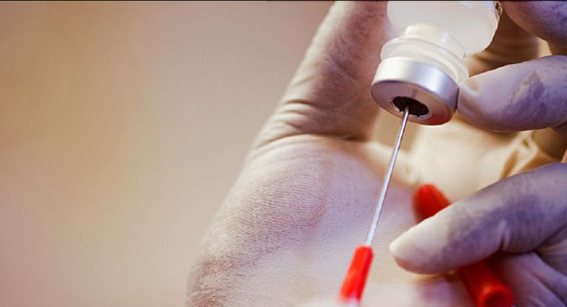
The Chinese team recruited women with breast cancer undergoing postoperative radiotherapy and randomized them (2:1) to receive either the green tea extract or placebo (normal saline solution). These solutions were sprayed to the whole of the radiation field from the first day of therapy until two weeks after completion of treatment.
A total of 165 women with a median age of 46 years were enrolled and randomized to EGCG, the primary catechin found in green tea or placebo.
The onset of radio-dermatitis was delayed by 2-3 weeks and the intensity and severity of the symptoms were significantly decreased in the treated group. No skin toxicity was observed.
The authors concluded that prophylactic use of a green tea extract significantly reduced both the incidence and severity of RID and that it has the potential to become a new choice for skin care in women receiving radiotherapy.
Topical green tea extract supports restoration of skin integrity and control of inflammatory cytokines and oxidative stress in the skin. Green tea extract also reduces the acute skin-induced reactions including pain and sensations of burning, itching, pulling and tenderness.
Dr. Chilkov: Practical Application:
 Topical Green Tea Extract Spray
To make a medicinal water extract: Place 8 organic green tea bags into a 16 oz glass jar or glass container. Pour boiling water over the tea bags, cover immediately and steep for one hour. After it has cooled to room temperature store covered in the refrigerator. When ready to use transfer water extract to a a glass spray bottle. Apply liberally to the radiation field before and after each radiotherapy session and three times daily for 3 weeks after the last radiotherapy session.
Fresh Aloe Vera Gel poultice
Areas where skin is most impacted can be covered with. mashed fresh aloe vera gel and covered with a large gauze bandage. This can easily be held in place underneath a sports bra or leotard or similar. Apply fresh aloe gel twice daily. Allow to be in contact with the skin for several hours or overnight. If you do not have access to a live aloe vera plant or fresh aloe gel you can use alcohol free aloe vera juice or aloe vera gel commonly found in natural foods stores. Aloe Vera is the botanical of choice for repair of radiation damaged skin.
Topical Calendula Oil (not extract) is also a soothing topical anti-inflammatory agent for radiation induced dermatitis. If the skin is very damaged, saturate a 4x4” gauze square and place over the affected area.
References:
Zhao H et al.
Efficacy of Epigallocatechin-3-Gallate (EGCG)in Preventing Dermatitis in Patients With Breast Cancer Receiving Postoperative Radiotherapy: A Double-Blind, Placebo-Controlled, Phase 2 Randomized Clinical Trial JAMA Dermatol 2022
Zhao H, et al.
Phase I study of topical epigallocatechin-3-gallate (EGCG) in patients with breast cancer
receiving adjuvant radiotherapy. Br J Radiol 2016; 89: 20150665.
Kyle T. Amber, BS et al
The Use of Antioxidants in Radiotherapy-Induced Skin Toxicity
Integrative Cancer Therapies 2014, Vol. 13(1) 38–45