Home » Nutritional Supplements

Category: Nutritional Supplements

green-tea-extract

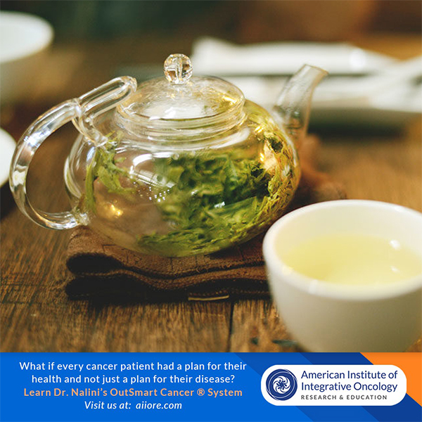
Green Tea Extract Reduces Severity of Radiation Dermatitis

The use of a solution containing a green tea extract has been shown to reduce both the incidence and severity of radiation-induced dermatitis in women undergoing adjunctive radiotherapy for breast cancer. This was the conclusion of a phase 2 randomized, placebo-controlled trial by a team of Chinese researchers.

Data from the World Health Organization indicates that in 2020, there were 2.3 million women diagnosed with breast cancer. In the treatment of women with breast cancer, radiation therapy is widely used conjunction with other therapies such as surgery, chemotherapy and hormonal therapies.  A common and frequent adverse effect of radiotherapy is radiation-induced dermatitis (RID) suffered by millions of women.

green-tea

The purpose of the current  study was to investigate the safety, tolerability and preliminary effectiveness of topical epigallocatechin-3-gallate (EGCG) for radiation dermatitis in patients with breast cancer receiving adjuvant radiotherapy.

A  solution of green tea extract  sprayed on the radiated areas of the skin reduced severity of radiation-induced dermatitis.

The Chinese team recruited women with breast cancer undergoing postoperative radiotherapy and randomized them (2:1) to receive either the green tea extract or placebo (normal saline solution).  These solutions were sprayed to the whole of the radiation field from the first day of therapy until two weeks after completion of treatment. 

A total of 165 women with a median age of 46 years were enrolled and randomized to EGCG, the primary catechin found in green tea or placebo.

The onset of radio-dermatitis was delayed by 2-3 weeks and the intensity and severity of the symptoms were significantly decreased in the treated group.  No skin toxicity was observed.

The authors concluded that prophylactic use of a green tea extract significantly reduced both the incidence and severity of RID and that it has the potential to become a new choice for skin care in women receiving radiotherapy.

Topical green tea extract supports restoration of skin integrity and control of inflammatory cytokines and oxidative stress in the skin. Green tea extract also reduces the acute skin-induced reactions including pain and sensations of burning, itching, pulling and tenderness.

Dr. Chilkov: Practical Application:

green-tea-leavesTopical Green Tea Extract Spray

To make a medicinal water extract: Place 8 organic green tea bags into a 16 oz glass jar or glass container.  Pour boiling water over the tea bags, cover immediately and steep for one hour.   After it has cooled to room temperature store covered in the refrigerator.  When ready to use transfer water extract to a a glass spray bottle.  Apply liberally to the radiation field before and after each radiotherapy session and three times daily for 3 weeks after the last radiotherapy session.  

Fresh Aloe Vera Gel poultice

Areas where skin is most impacted can be covered with. mashed fresh aloe vera gel and covered with a large gauze bandage.   This can easily be held in place underneath a sports bra or leotard or similar.  Apply fresh aloe gel twice daily. Allow to be in contact with the skin for several hours or overnight.  If you do not have access to a live aloe vera plant or fresh aloe gel you can use alcohol free aloe vera juice or aloe vera gel commonly found in natural foods stores.    Aloe Vera is the botanical of choice for repair of radiation damaged skin.

Topical Calendula Oil (not extract) is also a soothing topical anti-inflammatory agent for radiation induced dermatitis.  If the skin is very damaged, saturate a 4x4” gauze square and place over the affected area.

References:

Zhao H et al. 

Efficacy of Epigallocatechin-3-Gallate (EGCG)in Preventing Dermatitis in Patients With Breast Cancer Receiving Postoperative Radiotherapy: A Double-Blind, Placebo-Controlled, Phase 2 Randomized Clinical Trial JAMA Dermatol 2022

Zhao H, et al. 

Phase I study of topical epigallocatechin-3-gallate (EGCG) in patients with breast cancer

receiving adjuvant radiotherapy. Br J Radiol 2016; 89: 20150665.

Kyle T. Amber, BS et al

The Use of Antioxidants in Radiotherapy-Induced Skin Toxicity 

Integrative Cancer Therapies 2014, Vol. 13(1) 38–45

Probiotics

Oral Probiotics Reduce Complications of Surgery


Using probiotics before surgery prepares the patient for post operative stressors and complications. Using probiotics after surgery continues the support for the microbiome post operatively.

It is my practice to administer oral probiotics both before and after surgery with all of my patients.

Overall, using probiotics as part of pre-op and post-op care offers the following benefits

  • Reduction in Pro-Inflammatory Cytokines
  • Prevention of Surgical Infection and Sepsis 
  • Promotion of gastrointestinal microbial balance
  • Amelioration of adverse effects of oral antibiotics 
  • Decrease in adverse effects of opioids on gastrointestinal function
  • Promotion of Wound Healing at the surgical site

Use of oral probiotics is well tolerated and safe for use not only in cancer related surgeries but in a wide range of surgical procedures. 

Researchers conducting a randomized double blind placebo controlled study on the post operative effects of oral probiotics in patients undergoing resection for colorectal cancer concluded that probiotics not only decrease rates of infection at the incision site, respiratory and urinary systems but also inhibit proinflammatory factors such as TNFa, IL-17A , IL-17C, IL-22, IL-10 and IL-12.   Subjects in the treatment arm were given a 30 billion CFU mixture of six viable strains of Lactobacillus acidophilus, L. lactis, L. casei, Bifidobacterium longum, B. bifidum, and B. infantis twice daily for 6 months beginning 4 weeks postoperatively. [NB: I recommend starting pre-operatively].   Subjects in this arm did not experience infection, diarrhea or require antibiotics.

Zaharuddin L, Mokhtar NM, Muhammad Nawawi KN, Raja Ali RA. A randomized double-blind placebo-controlled trial of probiotics in post-surgical colorectal cancer. BMC Gastroenterol. 2019 Jul 24;19(1):131. doi: 10.1186/s12876-019-1047-4. PMID: 31340751; PMCID: PMC6657028.

In another study of patients receiving abdominal surgeries  oral probiotics were administered for 8 weeks.  The strains included were  L. plantarum, L. lactis, and L. delbrueckii. The study found statistically significant postoperative treatment reductions in abdominal pain and bloating, and significant improvements in stool formation. No clinically relevant adverse events were reported, and the treatment was well-tolerated by all patients. 

Bonavina L, Arini A, Ficano L, Iannuzziello D, Pasquale L, Aragona SE, Ciprandi G, On Digestive Disorders ISG. Post-surgical intestinal dysbiosis: use of an innovative mixture (Lactobacillus plantarum LP01, Lactobacillus lactis subspecies cremoris LLC02, Lactobacillus delbrueckii LDD01). Acta Biomed. 2019 Jul 10;90(7-S):18-23. doi: 10.23750/abm.v90i7-S.8651. PMID: 31292422; PMCID: PMC6776165.

In a recent 2021 Review of 14 studies of patients receiving gastrointestinal surgeries, a disruption of intestinal microbiome is identified and the prevalence of specific bacteria had significantly changed after surgery.

Ferrie S, Webster A, Wu B, Tan C, Carey S. Gastrointestinal surgery and the gut microbiome: a systematic literature review. Eur J Clin Nutr. 2021 Jan;75(1):12-25. doi: 10.1038/s41430-020-0681-9. Epub 2020 Jul 13. PMID: 32661352.

Another Review of 10 studies also identified post operative changes in the composition of the intestinal microbiome in patients receiving gastrointestinal surgeries  and posits that complications after gastrointestinal surgeries are linked to changes in the composition of the gut flora.

Lederer, A. K., Pisarski, P., Kousoulas, L., Fichtner-Feigl, S., Hess, C., & Huber, R. (2017). Postoperative changes of the microbiome: are surgical complications related to the gut flora? A systematic review. BMC surgery, 17(1), 125. https://doi.org/10.1186/s12893-017-0325-8

A study on the use of specific probiotics in patients undergoing resection for  colorectal cancer concluded that inflammatory cytokines and serum zonulin levels significantly decreased with probiotics. Probiotic ingestion resulted in compositional changes in gut microbiota; greater increases and decreases in healthy vs pathogenic bacteria, respectively, occurred with probiotics. Compositional increase in healthy bacteria was associated with reduced white blood cells, neutrophils, neutrophil-lymphocyte ratio, and zonulin. Bifidobacterium composition was negatively correlated with zonulin levels in the probiotic group, indicating repair of intestinal epithelium as an effective barrier. Probiotics improved postoperative flatus control and modified postoperative changes in microbiota and inflammatory markers.   In this study oral probiotics were administered both pre-op and post-op.  Probiotic supplementation included a mixture of three probiotic strains (Bifidobacterium animalis subsp. lactis HY8002 (1 × 10⁸ cfu), Lactobacillus casei HY2782 (5 × 10⁷ cfu), and Lactobacillus plantarum HY7712 (5 × 10⁷ cfu)

Park, I. J., Lee, J. H., Kye, B. H., Oh, H. K., Cho, Y. B., Kim, Y. T., Kim, J. Y., Sung, N. Y., Kang, S. B., Seo, J. M., Sim, J. H., Lee, J. L., & Lee, I. K. (2020). Effects of PrObiotics on the Symptoms and Surgical ouTComes after Anterior REsection of Colon Cancer (POSTCARE): A Randomized, Double-Blind, Placebo-Controlled Trial. Journal of clinical medicine, 9(7), 2181. https://doi.org/10.3390/jcm9072181

fight-prostate-cancer-vegetables

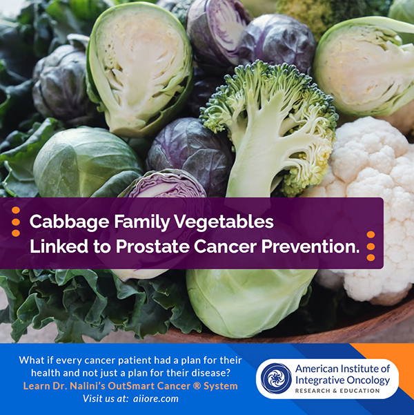
Prostate Cancer Chemoprevention: I3C and DIM

Increasing serum levels of phytochemical DIM may be chemopreventive and chemoprotective for prostate cancer.

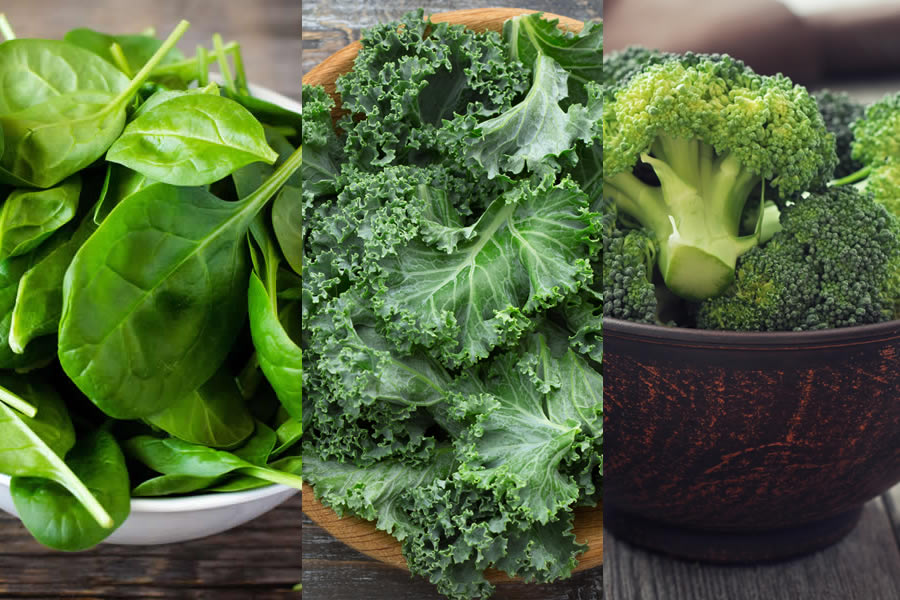
Cruciferous Cabbage Family vegetables such as (including cabbage, cauliflower, Brussels sprouts, broccoli, kale, arugula, bok choy and more ) are rich in dietary phytochemicals including Sulforaphane-Glucosinolate family molecules 13C (Indole-3-Carbinol) and it’s major bioactive therapeutic metabolite DIM  (3 3’di-indole methane).

Chopping, chewing, massaging and lightly steaming cruciferous vegetables activates the plants own catalyzing myrosinase enzyme and exposing the plant to the acid environment in the stomach leading to further metabolism resulting in bio available and bio active DIM.  DIM levels can be measured in the serum.

There are numerous studies on the benefits of dietary consumption of cruciferous vegetables.. A high intake of cruciferous vegetables is associated with reduced risk of several human cancers.  There are strong associations between high intake of broccoli and breast cancer and prostate cancer in humans.  Human cell studies show inhibition of  cell growth of several cancers including breast, prostate, pancreatic, colorectal, lung and head and neck cancers.

DIM and Prostate Cancer

DIM

DIM appears to be a potent inhibitor of human androgen hormones which may promote expression of androgen receptors on prostate cancer cells which may lead to carcinogenesis. Increasing serum levels of DIM through diet and supplementation may therefore be chemopreventive for prostate cancer.

Human and Cell Studies have shown that increased serum levels of DIM 

  • Reduces Serum Prostate Specific Antigen
  • Reduces Serum Androgen Hormones
  • Down Regulates Prostate Stem Cell Activity
  • Decreases  Nuclear Androgen Receptors
  • Induces p450 metabolic detoxification enzymes CYP1A1, CYP1A2 and CYP1B
  • Decreases oxidative stress via nrf2-KEAP pathway

The OutSmart Cancer® System is focused upon transforming the tumor microenvironment  which is a signaling environment, so that there is less physiologic support for the development and spread of cancer.   In a health model (rather than a disease model) we endeavor to transform the biosystem and  reduce pro-carcinogenic and proliferative signaling and prevent cellular, nuclear and mitochondrial damage.

brocolli

Therefore we use phytochemicals such as I3C and DIM and dietary interventions to influence carcinogenic and proliferative signaling in the tumor microenvironment.

Guidelines for Increasing Serum DIM levels

Primary Reference

*Amare DE. Anti-Cancer and Other Biological Effects of a Dietary Compound 3,3ʹ-Diindolylmethane Supplementation: A Systematic Review of Human Clinical Trials. Nutrition and Dietary Supplements. 2020;12:123-137

https://doi.org/10.2147/NDS.S261577

Additional Selected References 

  1. Anderton MJ, Manson MM, Verschoyle RD, et al. Pharmacokinetics and tissue disposition of indole-3-carbinol and its acid condensation products after oral administration to mice. Clin Cancer Res. 2004;10(15):5233–5241. doi:10.1158/1078-0432.CCR-04-0163
  2. Bjeldanes LF, Kim JY, Grose KR, et al. Aromatic hydrocarbon responsiveness-receptor agonists generated from indole-3-carbinol in vitro and in vivo: comparisons with 2, 3, 7, 8-tetrachlorodibenzo-p-dioxin. Proc Natl Acad Sci U S A. 1991;88(21):9543–9547. doi:10.1073/pnas.88.21.9543
  3. Chang Y-C, Riby J, Chang GH-F, et al. Cytostatic and antiestrogenic effects of 2-(indol-3-ylmethyl)-3, 3′-diindolylmethane, a major in vivo product of dietary indole-3-carbinol. Biochem Pharmacol. 1999;58(5):825–834. doi:10.1016/S0006-2952(99)00165-3
  4. Chen I, McDougal A, Wang F, et al. Aryl hydrocarbon receptor-mediated antiestrogenic and antitumorigenic activity of diindolylmethane. Carcinogenesis. 1998;19(9):1631–1639. doi:10.1093/carcin/19.9.1631
  5. Bradfield CA, Bjeldanes LF. High-performance liquid chromatographic analysis of anticarcinogenic indoles in Brassica oleracea. J Agric Food Chem. 1987;35(1):46–49. doi:10.1021/jf00073a010
  6. Weng J-R, Tsai C-H, Kulp SK, et al. Indole-3-carbinol as a chemopreventive and anti-cancer agent. Cancer Lett. 2008;262(2):153–163. doi:10.1016/j.canlet.2008.01.033
  7. Bradlow HL. Indole-3-carbinol as a chemoprotective agent in breast and prostate cancer. In Vivo. 2008;22(4):441–445
  8. Ahmad A, Ali S, Wang Z, et al. 3, 3′‐diindolylmethane enhances taxotere‐induced growth inhibition of breast cancer cells through downregulation of FoxM1. Int J Cancer. 2011;129(7):1781–1791. doi:10.1002/ijc.25839
  9. Ali S, Banerjee S, Ahmad A, et al. Apoptosis-inducing effect of erlotinib is potentiated by 3,3ʹ-diindolylmethane in vitro and in vivo using an orthotopic model of pancreatic cancer. Mol Cancer Ther. 2008;7(6):1708–1719. doi:10.1158/1535-7163.MCT-08-0354
  10. Banerjee S, Wang Z, Kong D, et al. 3, 3′-Diindolylmethane enhances chemosensitivity of multiple chemotherapeutic agents in pancreatic cancer. Cancer Res. 2009;69(13):5592–5600. doi:10.1158/0008-5472.CAN-09-0838
  11. Giovannucci E, Rimm EB, Liu Y, et al. A prospective study of cruciferous vegetables and prostate cancer. Cancer Epidemiol Prev Biomarkers. 2003;12(12):1403–1409.
  12. Kong D, Heath E, Chen W, et al. Loss of let-7 up-regulates EZH2 in prostate cancer consistent with the acquisition of cancer stem cell signatures that are attenuated by BR-DIM. PLoS One. 2012;7(3):e33729. doi:10.1371/journal.pone.0033729
  13. Abdelbaqi K,Lack N, Guns ET, et al. Antiandrogenic and growth inhibitory effects of ring‐substituted analogs of 3, 3′‐diindolylmethane (Ring‐DIMs) in hormone‐responsive LNCaP human prostate cancer cells. Prostate. 2011;71(13):1401–1412.
  14. Bhattacharjee S, Dashwood RH. Epigenetic Regulation of NRF2/KEAP1 by Phytochemicals. Antioxidants. 2020; 9(9):865. https://doi.org/10.3390/antiox9090865
  15. Li Y et al. Recent progress on nutraceutical research in prostate cancer. Cancer Metastasis Rev. 2014;33(2-3):629-640.
omega 3 fatty acids

Omega 3 Fatty Acids: Enhanced Control of Cancer Risk and Progression

A diet high in polyunsaturated fatty acids, especially omega 3s, have been shown to be negatively associated with cancer development

 Dietary fatty acids have been recognized as influential factors in the activation of carcinogenic events or disease progression and have been associated with a direct connection to breast cancer prevention.

PUFAs differentially inhibit mammary tumor development by inflicting modifications to the morphology of cell membranes, and influencing signaling pathways, gene expression and apoptosis.

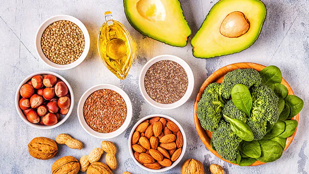
The human body is unable to synthesize long-chain polyunsaturated fatty acids (PUFAs) Omega 3 DHA, docosahexaenoic, and EPA, Eicosapentaenoic acid and Omega 6 Arachidonic Acid at a reasonable rate and therefore, supplementation is required through dietary sources or nutritional supplements. The recommended daily nutritional dose is 2,000 mg EPA+DHA, while therapeutic dosing is 4,000-6,000 milligrams of EPA+DHA per day.

omega-3-natural

 Omega Three Fatty Acids and the Tumor Microenvironment

  1. Supports Normal Inflammation Control by lowering COX 2, LOX5, PGE2, IL1, IL6,TNFa, CRP.
    • Increased inflammation contributes to cancer development, progression and metastasis.
    • Increased inflammation is linked to cancer related pain, fatigue, depression and cognitive impairment.
    • Increased inflammation is linked to cancer related hypercoagulation and risk of thromboembolism
    • Supporting Normal Inflammation control has a wide impact on the behavior of tumor cells and on safety and quality of life for cancer patients and survivors.
  2. Promotes Expression of M1 Type Tumor Associated Macrophages (TAMs).
    • Type M1 TAMs promote tumor regression, inflammation control and immune activation by promoting tumor infiltration by antigen presenting dendritic cells and cytotoxic T cells.
  3. Inhibits VEGF (Vascular Endothelial Growth Factor) and Promotes Normal Control of Angiogenesis .
    • VEGF promotes the development of new blood vessels to the tumor cells. Inhibition of VEGF and the development of capillaries inhibits tumor growth and profession as well as metastasis.
       
  4. Down regulates tumor promoter Protein Kinase C isoenzymes,
    • A group of enzymes that link multiple cellular processes responsible for regulation of tumorigenesis, cell cycle progression and metastasis.
  5. Inhibits Collagenase,
    • A proteolytic enzyme that breaks down the ECM (Extracellular Matrix) and allows invasion of tumor cells into tissues and blood vessels, leading to progression, invasion and metastasis.
  6. Promotes Normal Apoptosis signaling.
    • Cancer cells lose the ability to initiate apoptosis, the normal process in which a cell recognizes itself as aberrant and self destructs. The inhibition of normal apoptotic signaling in malignant cells is a hallmark  of the tumor microenvironment permissive of uncontrolled growth, persistence and immortality due to loss of normal regulation.
  7. Lowers Bcl2 and Ras oncogenes.
    • These genes inhibit normal apoptosis and promote tumor growth and progression.
  8. Acts as a Chemo-sensitizer
    • Working synergistically to enhance therapeutic effect of chemotherapy drugs. DHA has a potential to specifically chemo-sensitize tumors.
    • Tumour cells can be made more sensitive to chemotherapy than non-tumor cell when membrane lipids are enriched with DHA
    • Incorporating DHA during treatment reduces adverse effects of chemotherapy.
    • DHA can improve the outcome of chemotherapy when highly incorporated into cell membranes.
  9. Acts as a Radio-sensitizer.
    • By promoting normal membrane structure and function and by influencing the tumor microenvironment DHA acts synergistically to potentiate therapeutic effects of radiotherapy on tumor cells.
  10. Promotes Healthy 16-OH Estrogen metabolism.
    • Estrogen can be metabolized through multiple pathways. The promotion of 16-Hydroxylation of estrogen produces estrogen metabolites that are not pro-carcinogenic. Omega 3 Fatty Acids promote healthy estrogen metabolism.
  11. Inhibits Platelet Aggregation and Thrombin Formation.
    • Abnormal hyper-coagulation, increased platelet aggregation and thrombus formation are hallmarks of the tumor microenvironment. Control of platelet aggregation and thrombus formation reduces the risk of life threatening and adverse  thrombotic events.  40% of all cancer patients are at risk for the formation of thromboembolisms.  Omega 3 Fatty Acids reduce this risk.
  12. Promotes Normal Cell Membrane Functions and Receptor Binding
    • A healthy flexible cell membrane built of omega 3 fatty acids promotes an enhancement of all membrane functions, normalizing and optimizing normal and therapeutic physiology.
  13. Increases expression of Tumor Suppressor Gene PTEN.
    • Increased expression of tumor suppressor genes leads to enhanced control over carcinogenesis,  tumorigenesis and metastatic progression.
  14. Inhibits Multi Drug Resistance.
    • Tumor cells can quickly become resistant to therapeutic anti-neoplastic agents thus decreasing and shortening the efficacy of treatments.
  15. Inhibits cachexia preserves muscle mass and bone mass (inhibits proteolysis inducing factor)
    • Loss of bone mass (osteopenia) and loss of muscle mass (sarcopenia) are risk factors of aging and of the cancer physiology.  Maintaining bone mass and muscle mass are crucial to robust healthy function and quality of life.
  16. Supports normal mood regulation.
    • Depression and anxiety are common in cancer patients. Support of balanced mood allows cancer patients deep and restful sleep, improved quality of life and increased coping capacity and resilience in the face of stress.

Cautions and Contraindications

  • Patient on anticoagulant medications
  • Patients with thrombocytopenia and known hypo-coagultion clotting disorders
  • Pre and Post Surgical patients (72 hours)
  • Patients with seafood allergies


How to Measure Omega 3 Fatty Acid Status

Serum or Plasma Omega 3 Fatty Acid ratios. LABCORP Omega 3-6 Fatty Acids, Quest Diagnostics Omegacheck, Boston HeartLab Fatty Acid Balance, Cleveland HeartLab Omegacheck, Genova Diagnostics Essential and Metabolic Fatty Acids, Great Plains Comprehensive Fatty Acids, OmegaQuant Omega3 Index.

Selected References

 Azrad M, Turgeon C, Demark-Wahnefried W. Current evidence linking polyunsaturated Fatty acids with cancer risk and progressionFront Oncol. (2013) 3:224.

 Bartsch H, Nair J, Owen RW. Dietary polyunsaturated fatty acids and cancers of the breast and colorectum: emerging evidence for their role as risk modifiers. Carcinogenesis. (1999) 20:2209–18.

 Bournoux, P. Et al. Improving outcome of chemotherapy of metastatic breast cancer by DHA: Phase II Trial, Br.J Cancer 2009 Dec 15:101(12):1978-85

 Shweta Tiwary   Altered Lipid Tumor Environment and Its Potential Effects on NKT Cell Function and Tumor Immunity.  Front Immunol.10.3389/fimmu.2019.02187

 Zanoaga O, Jurj A, Raduly L, Cojocneanu-Petric R, Fuentes-Mattei E, Wu O, et al. Implications of dietary omega-3 and omega-6 polyunsaturated fatty acids in breast cancer.  Exp Ther Med. (2018) 15:1167–76. 10.3892/etm.2017.5515