Home » Uncategorized

Category: Uncategorized

Botanical and Phytochemical Radiosensitizers in Cancer Radiotherapy:

Integrating Natural Compounds for Optimal Clinical Outcomes

By Dr. Nalini Chilkov, L.Ac., O.M.D.

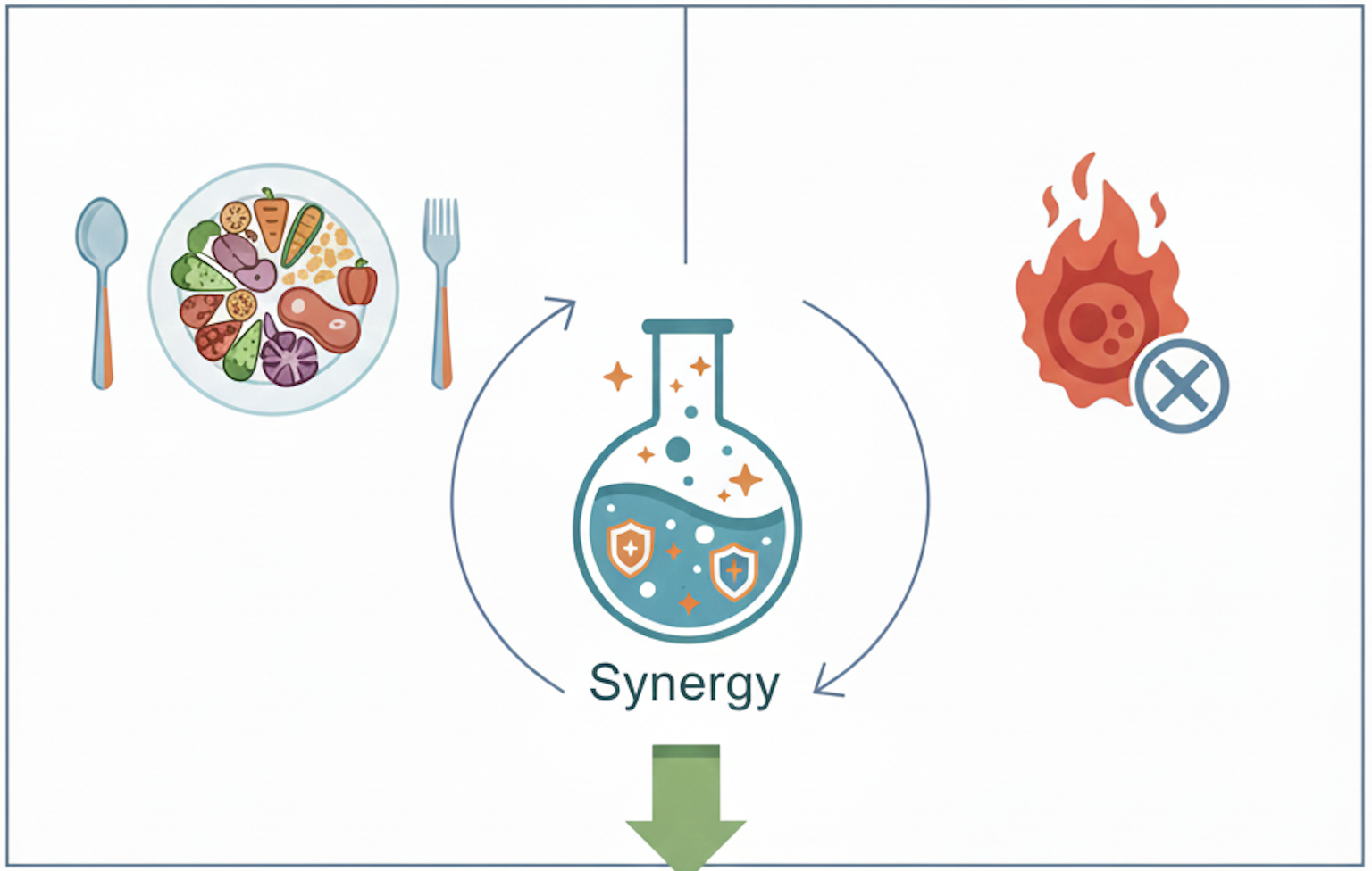
Botanicals uniquely offer dual synergist benefits: enhancing radiation-induced tumor cell death while simultaneously protecting healthy tissue, thereby maximizing therapeutic gain.

Integrating botanicals and phytochemicals as radiosensitizers provides a promising strategy to enhance therapeutic efficacy, reduce treatment side effects, and ultimately improve patient outcomes.

Integrating botanicals is not simply about enhancing radiosensitivity; it's about establishing a comprehensive health plan, creating a body where cancer cannot thrive, and reducing the anxiety and overwhelm that cancer patients often experience.

Phytochemicals as Powerful Radiosensitizers

Emerging evidence strongly supports integrating phytochemicals derived from food and medicinal plants into radiotherapy protocols:

Prominent phytochemicals and botanical sources include:
  1. Taxane diterpene (Taxus brevifolia – Pacific yew bark)
    Clinical trials confirm its radiosensitizing properties, especially in breast and ovarian cancers, by stabilizing microtubules and arresting cells in radiosensitive phases.² 
  2. Curcuminoids (Curcuma longa – Turmeric rhizome)
    Exhibits extensive radiosensitizing potential through NF‑κB and COX‑2 inhibition, enhanced ROS generation, and apoptosis induction. Undergoing clinical trials for glioblastoma, breast, and prostate cancers.² 
  3. Genistein Isoflavone (Glycine max – Soybeans)
    Enhances radiosensitivity by disrupting DNA repair, inducing G2/M arrest, and decreasing inflammation via NF‑κB inhibition. Clinical studies show reduction in radiation-induced fibrosis.² 
  4. Resveratrol stilbene (Vitis vinifera – Grapes, berries)
    Demonstrates potent radiosensitizing activity by amplifying ROS-mediated DNA damage and modulating PI3K/AKT/NF‑κB signaling pathways.² 
  5. Quercetin flavonoid (Allium cepa, Malus domestica – Onions, apples)
    Shows promise in vitro and in vivo by enhancing ROS production, causing cell cycle arrest, and protecting normal tissues from radiation injury.³ 
  6. Ellagic Acid polyphenol(Punica granatum, Rubus spp. – Pomegranates, berries)
    Preclinical studies reveal increased tumor cell death through elevated ROS and DNA damage.⁴ 
  7. Baicalein flavonoid (Scutellaria baicalensis – Chinese skullcap root)
    Nanoformulation demonstrates selective radiosensitization in breast cancer cells with limited toxicity to normal tissues.⁵ 
  8. Triphala polyphenols, flavonoids, phenolic acids, tannins, saponins, anthraquinones (Terminalia chebula, Terminalia bellerica, Emblica officinalis – Ayurvedic triplet)
    Exhibits tumor-selective radiosensitization and ROS elevation while sparing normal hepatocytes, beneficial for supportive cancer care.⁶ 
Traditional Chinese Medicine (TCM) Herbs as Radiosensitizers

Traditional Chinese Medicine (TCM) herbs have been employed therapeutically for millennia. Modern research confirms their potent radiosensitizing effects, primarily through apoptotic and autophagic pathways. Clinical and preclinical evidence has identified several effective TCM herbs:

  • Tanshinone IIA (Salvia miltiorrhiza – Danshen root): Induces G2/M cell cycle arrest and apoptosis, inhibits tumor metastasis via suppression of matrix metalloproteinases (MMP-2 and MMP-9), particularly in cervical and liver cancer models.¹ 
  • Ganoderma lucidum (Reishi mushroom): Demonstrates potent immunomodulatory and radioprotective properties, improving patient tolerance and recovery post-radiotherapy.¹ (caution using  immunostimulation polysaccharide rich Chinese Medicinal Mushrooms with patients also receiving immunotherapy.  There is a risk of exacerbating adverse inflammatory effects.  On the other hand, a patient with poor immune competency may benefit from the support.  In the OutSmart Cancer® System all care decisions must be thoughtfully and highly individualized.)
Understanding Mechanisms of Radiosensitization

Radiosensitizers are agents that selectively enhance the susceptibility of cancer cells to ionizing radiation. Botanicals and phytochemicals have become particularly compelling radiosensitizers due to their multiple mechanisms of action, excellent safety profiles, and additional anticancer properties. These natural compounds act by:

  • Increasing reactive oxygen species (ROS) generation 
  • Inducing cell cycle arrest at radiosensitive phases 
  • Enhancing apoptosis (programmed cell death) 
  • Reducing DNA repair capacity within tumor cells 
  • Modulating inflammation and immune response
Conclusion

Integrating botanical and phytochemical radiosensitizers represents an example of synergistic integrative  cancer care.  Through careful selection and thoughtful implementation, clinicians can offer patients not just a plan for disease treatment, but an integrated roadmap to both improved outcomes and better health.

As practitioners of integrative cancer care, our goal remains clear: to provide the tools, knowledge, and research-based interventions needed to ensure our patients get well, stay well, and live vibrantly well beyond cancer.

By harnessing nature’s pharmacy, the OutSmart Cancer® System can  leverage powerful synergies between conventional treatments and botanical medicine, transforming outcomes and improving the lives of cancer patients.

References (AMA Format):
  1. Jia L, Ma S, Hou X, et al. The synergistic effects of traditional Chinese herbs and radiotherapy for cancer treatment. Oncol Lett. 2013;5(5):1439-1447. doi:10.3892/ol.2013.1245 
  2. Komorowska D, Radzik T, Kalenik S, Rodacka A. Natural radiosensitizers in radiotherapy: Cancer treatment by combining ionizing radiation with resveratrol. Int J Mol Sci. 2022;23(18):10627. doi:10.3390/ijms231810627 
  3. Lin C, Yu Y, Zhao HG, Yang A, Yan H, Cui Y. Combination of quercetin and radiotherapy enhances tumor radiosensitivity in vitro and in vivo. Radiat Oncol. 2012;7:20. doi:10.1186/1748-717X-7-20 
  4. Ansari MA, Sinilal B, Anilakumar KR, Khan HA. Ellagic acid enhances the radiosensitivity of cancer cells through ROS-mediated DNA damage. Nutr Cancer. 2015;67(5):851-859. doi:10.1080/01635581.2015.1053494 
  5. Xiang H, Zhang L, Yang Z, Chen F, Zheng X, Cao H. Baicalein as a radiosensitizer for breast cancer: A nanoparticle approach. Int J Nanomedicine. 2021;16:2495-2508. doi:10.2147/IJN.S303846 
  6. Jagetia GC, Venkatesha VA. Enhancement of radiation effect by Triphala in tumor-bearing mice. Integr Cancer Ther. 2005;4(2):155-164. doi:10.1177/1534735405276448 
  7. Lawenda BD. Response to “Radiation Therapeutic Gain and Asian Botanicals,” by Stephen Sagar. Integr Cancer Ther. 2010;9(1):14-15. doi:10.1177/1534735410361476 
  8. Jelonek K, Musiał-Kulik M, Tomala J, et al. Prospective role of plant products in radiotherapy. Biomed Res Int. 2012;2012:363234. doi:10.1155/2012/363234 
  9. Garg AK, Buchholz TA, Aggarwal BB. Chemosensitization and radiosensitization of tumors by plant polyphenols. Antioxid Redox Signal. 2005;7(11-12):1630-1647. doi:10.1089/ars.2005.7.1630 
  10. Banerjee S, Singh SK, Chowdhury I, Lillard JW Jr, Singh R. Combinatorial effect of genistein and ionizing radiation suppresses prostate cancer growth in vivo. Cancer Res. 2005;65(11):4511-4518. doi:10.1158/0008-5472.CAN-05-0002 
  11. Ghafourian Boroujerdnia M, Khodagholi F, Emami SA, Mohammadirad A, Abdollahi M. Enhancement of radiotherapy by curcumin in glioblastoma cells: in vitro studies. Radiat Oncol. 2011;6:50. doi:10.1186/1748-717X-6-50 
  12. Smith TA, Kirkpatrick DR, Smith S, et al. Radioprotective agents to prevent cellular damage due to ionizing radiation. J Transl Med. 2017;15(1):232. doi:10.1186/s12967-017-1326-4 
  13. Tin AS, Sundar SN, Tran KQ, et al. Antiproliferative effects of Taccalonolide A from Tacca plantaginea: microtubule stabilizing properties distinct from paclitaxel. Biochem Pharmacol. 2008;76(4):482-493. doi:10.1016/j.bcp.2008.05.006 
  14. Sagar SM. Can the therapeutic gain of radiotherapy be increased by concurrent administration of Asian botanicals? Integr Cancer Ther. 2010;9(1):5-13. doi:10.1177/1534735409359772 


Social Isolation & Cancer: The Healing Power of Connection

As whole person care providers we must seek to understand our cancer patients’ ache of feeling physically or psychologically separated from loved ones, the toll of feeling unseen, unheard, or misunderstood. These wounds—of isolation, loneliness, and disconnection—are real; they affect the body/mind and spirit as profoundly as cancer treatments.  Today's research offers compelling evidence, and more importantly, meaningful tools we can use together to heal not just cancer, but its hidden burden: social isolation.

1. Why This Matters

A. Social Isolation Impacts Outcomes

B. Loneliness Deeply Affects the Mind

  • In survivors aged ≥50, those with high loneliness faced a 67% higher risk of death compared to those with minimal lonelinessfrontiersin.org+15oncologynurseadvisor.com+15jnccn.org+15.

  • Patients often fall silent under stress: fear of burdening others, the stigma of visible illness, or shame around changes in identity or body image. Studies link isolation with anxiety, depression, poor sleep, and negative self-image.

C. Biology & Behavior—How Isolation Impacts Health

  • Isolation intensifies inflammation, suppresses immune surveillance, and dysregulates cortisol and autonomic balance—all biologic stressors that may fuel tumor growth and impair treatment response.

  • Behaviorally, isolated patients are more likely to skip appointments, neglect medications, eat poorly, and withdraw from activity—all of which undermine recovery.

2. Caring Steps : Talk to Patients, Make Referrals

In my practice, I see four essential elements:

A. Gentle Screening

At key moments—diagnosis, treatment transitions, and follow-up—I ask open questions:

  • “How connected have you felt lately?”

  • “Do you often wish for someone to talk to?”
    Using brief tools like the UCLA Loneliness Scale or a simple social network check, I watch for signals that isolation may be causing you avoidable pain.

B. Compassionate Listening + Referrals

If loneliness is weighing on you, I invite you to share what’s behind it. Then:

  • Connect patients with  oncology social work team.

  • Refer  to psycho-oncology counselors skilled in therapies like CBT or Mindfulness-Based Stress Reduction—both shown to reduce loneliness and psychological distress.

C. Peer Connection

  • In-person peer groups (especially helpful for breast, colorectal, or lung cancer survivors) reduce anxiety, restore hope, and ease isolation.

  • Online communities and forums can connect patients with people who truly understand; they offer emotional reinforcement and practical tips, and studies show they boost treatment adherence and well-being.

  • Buddy systems or livestream peer meetups: simple but powerful — just a 30-minute weekly check-in can build resilience and normalize your experience.

D. Tailored Lifestyle & Community Integration

  • Encourage gentle group exercise (yoga, walking groups), which supports physical function and elevates moodverywellhealth.com.

  • Partner with local nonprofits—Cancer Support Community, Lance Armstrong Foundation, or metastatic breast cancer groups—to link you with trusted activities and meaningful companionship.

  • Promote spiritual or creative engagement—whether in nature, art, music, faith—to enhance connectedness and purpose.

3. A Compassionate Clinical Blueprint for Care Givers and Patients

Step What I Do What You Do Why It Matters
1️⃣ Screen Start the conversation with caring assessment Be honest about how you're feeling So I can see and not overlook your need
2️⃣ Listen & Refer Provide support and arrange referrals Attend support sessions or therapy To reconnect you to someone who understands
3️⃣ Engage Peer Support Introduce you to peer groups and online communities Join at least one group or buddy partnership Shared stories can heal isolation
4️⃣ Promote Activity & Community Prescribe gentle group activity; connect with local resources Try a new group or routine weekly Movement + community = nourishment
5️⃣ Monitor & Adapt Re-check loneliness and mental health periodically Share back what’s working and what's hard To fine-tune your support over time

4. A Story of Connection

Meet “Maria,” age 60, recovering from colorectal cancer surgery.
At first, she was stoic and quiet about her isolation. Gentle questions revealed she felt she “had no one who really gets it.” I referred Maria to a psychotherapist skilled in CBT and to a local colorectal cancer peer support group. Over weeks, she began a low-impact walking class with fellow survivors and joined an online forum. Her mood lifted. She slept better, showed up for her follow ups on time, and started gardening again—something she loved before. Three months later, she told me: “I thought I was alone in this fight. Now I know I’m not.”

5. What the Research Tells Us

6. Let Patients Know How They Can Take Action  

  1. Reach out. Tell me or a member of your care team when you’re feeling alone.
  2. Accept help. Let others in—whether it's family, peers, or professionals.
  3. Stay in motion. Join gentle group exercise or activity-based support.
  4. Log in. Explore online groups that feel safe, moderated, and aligned with your needs.
  5. Give Feedback. Check in during follow-ups: Is this working? What needs adjusting?

7. Final Reflections: Connect, Comfort, Inspire

Together, we can do more than treat cancer. We can heal loneliness, restore hope, and rebuild a sense of connecting and belonging—step by step, day by day.

Cancer is a journey, and the lonely road is much harder to walk. The science shows that isolation isn’t just painful—it’s dangerous. But the good news is that connection, community, and compassion change outcomes. They lower stress, boost immunity, improve adherence, and may even help prevent recurrence.

As your physician, I care about your scans—and your soul. I see your strength, but also the hard days. I’m here with you—not just in the clinic, but in calling in help, helping you rebuild your social world, and walking this path together.

References

  1. 1. Wang F, Gao Y, Han Z, et al. A systematic review and meta-analysis of 90 cohort studies of social isolation, loneliness and mortality. Nat Hum Behav. 2023;7(8):1307‑1319. doi:10.1038/s41562-023-01617-6 academic.oup.comen.wikipedia.org+7nature.com+7ideas.repec.org+7
  2. 2. Holt‑Lunstad J, Smith TB, Layton JB. Social relationships and mortality risk: a meta-analytic review. JAMA Netw Open. 2023;6(1):e236921. doi:10.1001/jamanetworkopen.2023.6921 en.wikipedia.org+1en.wikipedia.org+1
  3. 3. Oncology Nurse Advisor. Loneliness and isolation for cancer patients: risk and interventions. Oncology Nurse Advisor. 2025. bps.org.uk+4oncologynurseadvisor.com+4academic.oup.com+4
  4. 4. Cancer Phys Outcomes; retrospective longitudinal study of 3447 cancer survivors aged ≥50 found those with highest loneliness had HR = 1.67 for mortality. Oncology Nurse Advisor. 2024. thetimes.co.uk+2oncologynurseadvisor.com+2pressroom.cancer.org+2
  5. 5. Medical News Today. Social isolation, loneliness may raise mortality risk, study finds. Med News Today. 2023. sciencedirect.com+15medicalnewstoday.com+15jamanetwork.com+15
  6. 6. Nature. Loneliness among adult cancer survivors in the United States; estimated 35.9% experienced moderate-severe loneliness. Sci Rep. 2025;14:85126. nature.com
  7. 7. Mediation analysis study: social isolation–cancer mortality link is 74% mediated by health behavior. Health Psychol. 2021;40(1):e1‑e10. sciencedirect.com+1researchgate.net+1


Microbiome and Immunotherapy

Targeting the Gut Microbiome and Metabolic Pathways to Enhance Immunotherapy Outcomes in Cancer Care

By Dr. Nalini Chilkov, L.Ac., O.M.D.
Founder, OutSmart Cancer® System

Introduction

Immunotherapy has revolutionized the treatment landscape for many types of cancer. Yet, response rates remain variable and unpredictable. Immune checkpoint blockade (ICB) therapies are often undermined by tumor-induced immunosuppression, systemic inflammation, and adverse effects stemming from immune hyperactivation. Emerging evidence highlights the gut microbiome and metabolic environment as critical modulators of immunotherapy efficacy and tolerance.

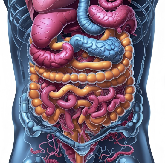
Response to immunotherapy  can be enhanced by optimizing  the health of the microbiome, supporting robust immunity, and promoting systemic metabolic health. These are fundamental elements of the OutSmart Cancer® System.

The Gut-Immune-Cancer Axis

The gut microbiome is now recognized as a powerful regulator of immunity, metabolism, and systemic inflammation. Composed of trillions of microorganisms, this ecosystem influences T-cell function, immune checkpoint pathways, and the tumor microenvironment (TME).

Key mechanisms include:

[Read More]

  • Modulation of T-cell activation and exhaustion.
  • Regulation of dendritic cell antigen presentation.
  • Production of immunomodulatory metabolites (e.g., short-chain fatty acids, indoles, inosine).
  • Maintenance of gut barrier integrity, reducing systemic endotoxemia.

Clinical studies show that high microbial diversity (α-diversity) and the presence of specific beneficial strains—such as Faecalibacterium prausnitzii, Akkermansia muciniphila, and Bifidobacterium longumare associated with improved response rates to immune checkpoint blockade and reduced risk of immune related adverse events6.

Conversely, gut dysbiosis—often caused by antibiotics, low-fiber diets, or systemic inflammation—can impair immune surveillance and reduce therapeutic benefit. Patients with Proteobacteria-dominant microbiomes show poorer outcomes and increased toxicity6.

Diet and the Microbiota-Immune Interface

The Mediterranean Diet: A Clinical Standard

The OutSmart Cancer Diet is a low glycemic modification of the well-studied Mediterranean Diet (MD) which emphasizes vegetables, legumes, whole grains, fruits, nuts, olive oil, and modest intake of fish and poultry. It has been shown to:

  • Increase SCFA-producing bacteria (e.g., Clostridium cluster XIVa, F. prausnitzii).
  • Reduce inflammation and enhance immune regulation.
  • Improve absorption of essential micronutrients (vitamin D, iron, folate).

The OutSmart Cancer® Diet reduces the carbohydrate intake of the Mediterranean Diet to achieve lower normal glucose and insulin levels to reduce the insulinogenic growth signaling and to support mitochondrial fatty acid rather than glucose dominant energy metabolism.

The OutSmart Cancer® Diet also encourages patients to include small portions of a variety of natural fermented foods in their daily diets as a source of a wide spectrum of healthy microorganisms to support a diverse and robust intestinal microbiome.

In a 2023 study published in JAMA Oncology, adherence to the MD was associated with significantly higher objective response rates and progression-free survival in advanced melanoma patients treated with ICB6.

High-Fiber Diets and Whole Plant Foods

Soluble dietary fiber from vegetables fuels beneficial microbes and promotes production of butyrate, a SCFA with anti-inflammatory and immune-supportive properties. High-fiber diets:

  • Increase microbial diversity.
  • Decrease gut permeability and systemic LPS levels.
  • Support regulatory T-cell activity and mucosal immunity.

A landmark 2021 Science study showed that patients with higher fiber intake and no probiotic use had significantly better responses to ICB (82% vs. 59%) and prolonged PFS6.

Metabolic Interventions: Fasting and Caloric Restriction

Cancer cells exhibit high metabolic demands and are less adaptable to nutrient scarcity. Modifying caloric intake alters host metabolism, immune cell dynamics, and the TME—shifting the balance in favor of immune activation and tumor suppression.

Short-Term Water Fasting (STWF)

STWF involves complete abstinence from food for 72–120 hours with maintained hydration. It:

  • Decreases IGF-1, insulin, and glucose levels.
  • Increases ketone bodies and autophagy.
  • Protects normal cells from chemotherapy-induced toxicity (differential stress resistance).
  • Enhances cytotoxic T-cell and NK cell activity1,2.

Fasting-Mimicking Diet (FMD)

The FMD replicates the benefits of fasting while providing limited caloric intake (plant-based, low-protein, high-fat) for 5 days. Clinical trials have demonstrated that FMD:

  • Lowers systemic inflammation and metabolic markers (glucose, IGF-1).
  • Depletes immunosuppressive cells (MDSCs, Tregs).
  • Enhances ICB efficacy and reduces irAEs in melanoma and NSCLC patients3.

Time-Restricted Eating (TRE)

TRE aligns eating patterns with circadian rhythms by confining caloric intake to 8–10 hours daily. Benefits include:

  • Improved insulin sensitivity, weight management, and lipid profiles.
  • Reduced inflammatory cytokines (TNF-α, IL-6).
  • Increased microbial diversity and butyrate-producing bacteria4,5.

Functional Nutrients and Immune Support

Vitamin D

Vitamin D (VD) modulates both innate and adaptive immunity. It enhances mucosal barrier integrity, reduces inflammatory cytokine production, and promotes regulatory T-cell function. VD deficiency is common in cancer patients and may:

  • Increase gut permeability and systemic inflammation.
  • Correlate with higher rates of irAEs.
  • Impair microbiome diversity6.

Omega-3 Fatty Acids

Omega-3 polyunsaturated fatty acids (PUFAs) such as EPA and DHA:

  • Suppress NF-κB activation and COX-derived pro-inflammatory mediators.
  • Increase SCFA-producing gut bacteria.
  • Are associated with improved B-cell and NK cell activity.

Safe dosing ranges from 1.5 to 3 g/day. These fatty acids are generally well-tolerated and help preserve lean body mass and immune tone6.

Clinical Integration: Actionable Strategies

Step 1: Dietary Guidance

  • Emphasize a low carb OutSmart Cancer® modified Mediterranean-style diet.
  • Encourage high-fiber, unprocessed plant foods.
  • Minimize processed meat, alcohol, and refined sugar.

Step 2: Fasting Protocols

  • STWF (72 hours) or FMD (5-day cycles) in supervised settings.
  • TRE (8–10 hour eating window) as a sustainable, daily intervention.

Step 3: Supplement Wisely

  • Test and correct vitamin D insufficiency.
  • Supplement omega-3s if inflammatory burden or cachexia is present.

Step 4: Preserve the Microbiome

  • Avoid non-essential antibiotic use near ICB initiation.
  • Favor food-based prebiotics over commercial probiotics unless indicated.

Step 5: Educate and Empower

  • Teach patients how lifestyle and nutrition influence outcomes.
  • Reinforce the message: Cancer is not in control. You are.

Conclusion

A paradigm shift is underway in oncology—one that embraces the whole patient, not just the tumor. By targeting the gut microbiome and metabolic pathways, we can optimize immunotherapy outcomes, reduce adverse effects, and empower patients to play an active role in their healing.

Create a body where cancer cannot thrive.
Support the Tumor Microenvironment. Strengthen the immune system. Transform outcomes.

Bibliography

  1. Brandhorst S., Longo V. D. (2019). Fasting and fasting-mimicking diets for chemotherapy augmentation. GeroScience, 41(4), 387–398. https://doi.org/10.1007/s11357-019-00072-5
  2. Dorff T. B., et al. (2016). Safety and feasibility of fasting in combination with platinum-based chemotherapy. BMC Cancer, 16, 360. https://doi.org/10.1186/s12885-016-2370-6
  3. Vernieri C., et al. (2022). Fasting-mimicking diet is safe and reshapes metabolism and antitumor immunity in patients with cancer. Cancer Discovery, 12(1), 90–107. https://doi.org/10.1158/2159-8290.CD-21-0030
  4. Wilkinson M. J., et al. (2020). Ten-hour time-restricted eating reduces weight, blood pressure, and atherogenic lipids in patients with metabolic syndrome. Cell Metabolism, 31(1), 92–104.e5. https://doi.org/10.1016/j.cmet.2019.11.004
  5. Di Francesco A., et al. (2018). A time to fast. Science, 362(6416), 770–775. https://doi.org/10.1126/science.aau2095
  6. David A., Lev-Ari S. (2024). Targeting the Gut Microbiome to Improve Immunotherapy Outcomes: A Review. Integrative Cancer Therapies, 23:1–10. https://doi.org/10.1177/15347354241269870


PART TWO Red Meat Consumption and Cancer Risk

Clinical Implications for Integrative Oncology Practice

As integrative oncology physicians, we are entrusted with guiding patients toward dietary patterns that support their health during and after cancer treatment. Understanding the role of red and processed meats in the cancer terrain empowers us to educate patients about evidence-based choices that can influence cancer outcomes and survivorship.

Dietary Counseling and Cancer Prevention

In clinical practice, discussions about meat consumption should be personalized. Patients with a history of colorectal, breast, or prostate cancer may benefit from eliminating processed meats and limiting red meat intake. Encourage the adoption of dietary patterns rich in anti-inflammatory, antioxidant, and fiber-rich foods, such as the Mediterranean or plant-forward diets, which have been shown to support immune function and reduce chronic inflammation—key factors in the Cancer Terrain.

Key Recommendations for Physicians:

  • Assess Meat Intake: Routinely inquire about red and processed meat consumption during dietary assessments, especially for patients with known cancer risk factors or histories.
  • Promote Plant-Based Proteins: Recommend legumes, nuts, seeds, and organic tofu or tempeh as alternatives to red meat. These options provide adequate protein without the carcinogenic risks associated with processed meats. 
  • Educate on Cooking Methods: Advise patients to avoid high-temperature cooking methods that generate carcinogens. Recommend steaming, stewing, or baking at lower temperatures. 
  • Encourage Dietary Diversity: Promote a diet abundant in colorful vegetables, fruits, whole grains, and phytonutrient-rich herbs and spices to support detoxification pathways and healthy cellular function. 

A Broader Perspective on Risk Reduction

While no single food causes or cures cancer, dietary patterns can shape the Cancer Terrain—the internal environment that influences whether cancer will develop or progress. By reducing exposure to known dietary carcinogens and increasing intake of protective compounds, patients can take an active role in their healing journey.

Conclusion

The link between red and processed meat consumption and cancer risk is supported by robust evidence, particularly for colorectal and breast cancers. Integrative oncology physicians must translate this knowledge into actionable guidance, empowering patients to make informed dietary choices. This approach not only aligns with cancer prevention but also fosters overall health and vitality—goals that resonate deeply with our patients' aspirations to Get Well, Stay Well, and Live Well Beyond Cancer.

References

  1. IARC Monographs. Red Meat and Processed Meat. International Agency for Research on Cancer. https://www.iarc.who.int/wp-content/uploads/2018/07/pr240_E.pdf 
  2. Farvid MS, et al. Meat intake and risk of breast cancer: A systematic review and meta-analysis. Int J Cancer. 2021;149(8):1512-1524. https://pubmed.ncbi.nlm.nih.gov/34455534 
  3. Zhao Z, et al. Red and processed meat consumption and gastric cancer risk: A systematic review and meta-analysis. Oncotarget. 2017;8(18):30563-30575. https://pmc.ncbi.nlm.nih.gov/articles/PMC5444765 
  4. Lippi G, et al. Red meat consumption and cancer risk: A critical review. Crit Rev Food Sci Nutr. 2023. https://pmc.ncbi.nlm.nih.gov/articles/PMC10577092 
  5. Diallo A, et al. Red and processed meat intake and prostate cancer risk: A systematic review and meta-analysis of prospective studies. Nutrients. 2022;14(5):1036. https://pubmed.ncbi.nlm.nih.gov/35198587 
  6. IARC Monograph Volume 114. Red Meat and Processed Meat. WHO Publications. https://publications.iarc.who.int/Book-And-Report-Series/Iarc-Monographs-On-The-Identification-Of-Carcinogenic-Hazards-To-Humans/Red-Meat-And-Processed-Meat-2018 
  7. IARC Q&A. Carcinogenicity of consumption of red and processed meat. https://www.iarc.who.int/wp-content/uploads/2018/11/Monographs-QA_Vol114.pdf 

To your health, healing, and wholeness.
Create a Body Where Cancer Cannot Thrive.

Red Meat Consumption and Cancer Risk

PART ONE 

Red Meat Consumption and Cancer Risk 

​The relationship between red meat consumption and cancer risk has been extensively investigated. Epidemiological studies and meta-analyses have provided insights into how red meat and processed meat intake may influence the incidence of various cancers.​+5PMC+5PubMed+5

Several mechanisms are proposed to elucidate the correlation between the consumption of red and processed meat and the risk of cancer.  These mechanisms include:

  • Heme Iron: The high concentration of heme iron in red meat facilitates the formation of N-nitroso compounds, acknowledged carcinogens that may induce damage to the gastrointestinal lining.
  • Cooking Methods: The utilization of elevated-temperature cooking techniques, such as grilling and barbecuing, promotes the synthesis of heterocyclic aromatic amines and polycyclic aromatic hydrocarbons, which possess carcinogenic potential.
  • Processing Chemicals: Processed meats frequently incorporate nitrates and nitrites, which can undergo transformation into N-nitroso compounds during the digestive process, thereby contributing to carcinogenesis.

Cancers Linked To Red Meat Consumption

Colorectal Cancer

The International Agency for Research on Cancer (IARC) has classified processed meat as “carcinogenic to humans” (Group 1) and red meat as “probably carcinogenic to humans” (Group 2A), with a significant association observed between processed meat consumption and colorectal cancer. Specifically, an analysis estimated that every 50-gram portion of processed meat consumed daily increases the risk of colorectal cancer by about 18%. ​

Breast Cancer

A comprehensive systematic review and meta-analysis indicated that high red meat intake is positively associated with an increased risk of breast cancer. Similarly, high processed meat intake was linked to a higher risk of breast cancer. ​PubMed+2PubMed+2PMC+2

Gastric Cancer

The association between red and processed meat consumption and gastric cancer risk has yielded mixed results. Some case-control studies suggest a positive association, while cohort studies have not consistently supported this link. Therefore, the evidence remains inconclusive. ​PubMed+2PMC+2PMC+2PMC

Pancreatic Cancer

Research has indicated a potential link between high red meat consumption and an increased risk of pancreatic cancer. However, further studies are needed to confirm this association. ​IARC PublicationsPMC

Prostate Cancer

A systematic review and meta-analysis of prospective studies suggested that increased consumption of total meat and processed meat might be associated with a higher risk of prostate cancer. ​PMC+2PubMed+2PubMed+2

Conclusion

The body of evidence suggests a positive association between red and processed meat consumption and the risk of several cancers, notably colorectal and breast cancer. While the exact mechanisms remain under investigation, it is prudent for healthcare professionals to consider these findings when advising patients on dietary choices. All patients are advised to limit red meat consumption and eliminate all processed meats. The OutSmart Cancer® diet  recommends poultry fish, eggs and legumes as healthy sources of protein It is plant strong, rich in fruits, vegetables, and healthy fats may be supports a healthy and lower risk tumor microenvironment.

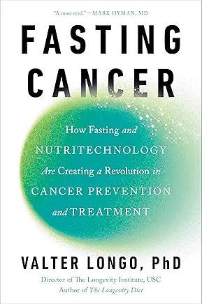
Dr. Valter Longo’s New Book: Fasting Cancer

Dr. Valter Longo, a prominent figure in the field of biogerontology, has extensively researched the intersection of nutrition, fasting, and cancer therapy. His recent work, particularly encapsulated in his 2025 publication, Fasting Cancer: How Fasting and Nutritechnology Are Creating a Revolution in Cancer Prevention and Treatment, provides a comprehensive overview of his findings and methodologies.

Key Insights from Fasting Cancer

In Fasting Cancer, Dr. Longo presents decades of research highlighting how controlled dietary interventions, specifically fasting and fasting-mimicking diets (FMDs), can enhance cancer treatment outcomes. He elucidates that while cancer cells are adept at exploiting various nutrients for growth, they become particularly vulnerable during fasting states. In contrast, healthy cells activate protective mechanisms under these conditions, leading to a differential stress response that can be therapeutically advantageous. ​

A notable quote from Dr. Longo emphasizes this concept:​

“While we have learned that you cannot starve cancer with fasting alone, since cancer steals from other cells and finds a way to stay alive even if the patient fasts, you can use fasting-mimicking diets to make the cancer cells so weak or desperate that the right therapy will kill them.”  

Furthermore, the book discusses how integrating his Longevity Diet—a plant-based nutritional regimen—and plant-based ketogenic diets can support and potentially amplify the efficacy of conventional cancer therapies. Dr. Longo underscores the importance of patients actively participating in their treatment plans through dietary strategies, aiming to make therapies more effective and less toxic

Recent Research Highlights

Dr. Longo's recent studies have provided empirical support for the benefits of FMDs in oncology:​

  1. Breast Cancer Study (2024): A sub-analysis of the NCT03340935 trial indicated that incorporating FMD cycles with first-line carboplatin-based chemotherapy was associated with improved overall survival in patients with advanced triple-negative breast cancer.
  2. Chronic Lymphocytic Leukemia (2024): Research demonstrated that cyclic FMD, when combined with Bortezomib and Rituximab, effectively treated chronic lymphocytic leukemia, suggesting a synergistic effect between FMD and these therapeutic agents. ​
  3. T-Cell Leukemia (2023): Findings revealed that FMD inhibited autophagy and, in combination with chemotherapy, promoted T-cell-dependent leukemia-free survival, highlighting the potential of FMD to enhance immune-mediated cancer clearance.

 These studies collectively suggest that FMDs can play a pivotal role in reprogramming cancer cell metabolism, improving the efficacy of standard treatments, and reducing adverse side effects.

PART 4: Intermittent Fasting: Considerations for Clinical Practice

Intermittent Fasting and Cancer Metabolism:   A Four Part Series

The OutSmart Cancer® System recommends intermittent fasting for 13 hours of calorie restriction per 24 hour cycle with the appropriate assessment, monitoring and guidance of a health care provider.

While the results are promising, it's important to note that more extensive clinical trials are needed to establish standardized guidelines for implementing IF in oncology.

Personalized approaches should be considered, as the effects of IF may vary depending on cancer type, stage, and individual patient factors.

 Therefore, personalized approaches, clinical supervision and monitoring and further research are needed to fully understand and optimize the use of IF in cancer treatment.

Intermittent fasting represents a multifaceted approach to cancer prevention and treatment, leveraging metabolic, immunological, and cellular mechanisms. As research in this field continues to evolve, IF may become an important adjunct to conventional cancer therapies and part of the OutSmart Cancer® System for Creating a

 For Part 5 simply publish the blog post but a newsletter does not have to be sent

Intermittent Fasting and Cancer Metabolism:  Selected References

Part 5: Intermittent Fasting: Selected References

  1. Anemoulis, et al. (2024). Intermittent Fasting on Cancer: An Update. EJ Clinical Medicine.
  2. de Groot, S., et al. (2022). Effect of fasting on cancer: A narrative review of scientific evidence. Nutrition Reviews, 80(10), 2159-2177.
  3. Clifton, K. D., et al. (2021). Intermittent fasting in the prevention and treatment of cancer. CA: A Cancer Journal for Clinicians, 71(6), 527-546.
  4. Ferro, K., et al. (2023). Impact of Fasting on Patients With Cancer: An Integrative Review. Clinical Journal of Oncology Nursing, 27(6), 619-625.
  5. Nencioni, A., et al. (2018). Fasting and cancer: molecular mechanisms and clinical application. Nature Reviews Cancer, 18(11), 707-719.
  6. Harvie, M., & Howell, A. (2023). Intermittent fasting interventions to leverage metabolic and circadian rhythms for cancer prevention and control. JNCI Monographs, 2023(61), 84-93.
  7. Morales-Suárez-Varela, M., et al. (2020). Effect of intermittent fasting on cancer prevention: a systematic review. European Journal of Public Health, 30(Supplement_5), ckaa166.216.
  1. Stringer, A. M., et al. (2018). Autophagy and intermittent fasting: the connection for cancer therapy? Clinics, 73(suppl 1), e814s.
  2. Longo, V. D., & Mattson, M. P. (2014). Fasting: molecular mechanisms and clinical applications. Cell metabolism, 19(2), 181-192.
  3. Brandhorst, S., & Longo, V. D. (2016). Fasting and caloric restriction in cancer prevention and treatment. Recent Results in Cancer Research, 207, 241-266.
  4. Antunes, F., et al. (2018). Autophagy and intermittent fasting: the connection for cancer therapy? Clinics, 73(suppl 1), e814s.
  5. Lee, C., & Longo, V. D. (2016). Fasting vs dietary restriction in cellular protection and cancer treatment: from model organisms to patients. Oncogene, 35(15), 1475-1490.
  6. Safdie, F. M., et al. (2009). Fasting and cancer treatment in humans: A case series report. Aging (Albany NY), 1(12), 988-1007.
  7. de Groot, S., et al. (2019). Fasting mimicking diet as an adjunct to neoadjuvant chemotherapy for breast cancer in the multicentre randomized phase 2 DIRECT trial. Nature communications, 11(1), 3083.
  8. Freedland, S. J., et al. (2024). Researchers Look to Fasting as a Next Step in Cancer Treatment. Cedars-Sinai Discoveries.
  9. Cheng, C. W., et al. (2014). Prolonged fasting reduces IGF-1/PKA to promote hematopoietic-stem-cell-based regeneration and reverse immunosuppression. Cell stem cell, 14(6), 810-823.
  10. Di Biase, S., et al. (2016). Fasting-mimicking diet reduces HO-1 to promote T cell-mediated tumor cytotoxicity. Cancer cell, 30(1), 136-146.
  11. Caffa, I., et al. (2020). Fasting-mimicking diet and hormone therapy induce breast cancer regression. Nature, 583(7817), 620-624.
  12. Wei, S., et al. (2017). Fasting-mimicking diet and markers/risk factors for aging, diabetes, cancer, and cardiovascular disease. Science translational medicine, 9(377), eaai8700.
  13. Buono, R., & Longo, V. D. (2018). Starvation, stress resistance, and cancer. Trends in Endocrinology & Metabolism, 29(4), 271-280.

PART 3: Intermittent Fasting and Insulin Like Growth Factor (IGF-1)

Intermittent Fasting and Cancer Metabolism:   A Four Part Series

PART 3: Intermittent Fasting and Insulin Like Growth Factor (IGF-1)


Intermittent fasting (IF) reduces insulin-like growth factor 1 (IGF-1) levels through several specific mechanisms:

  1. Protein Restriction: The reduction in IGF-1 is primarily attributed to the restriction of protein intake, particularly essential amino acids, during fasting periods8.

READ MORE

  1. Calorie Restriction: The decrease in overall calorie intake during fasting supports the reduction in IGF-1 levels8.
  2. Insulin Reduction: Fasting leads to a significant decrease in circulating insulin levels, which in turn promotes the reduction of IGF-182.
  3. Metabolic Switch: IF induces a metabolic switch that mimics water fasting, leading to a decrease in IGF-1 levels. For example, a 4-day fasting-mimicking diet (FMD) can reduce IGF-1 levels by approximately 45%3.
  4. IGFBP-1 Increase: Fasting causes a substantial increase in IGF binding protein 1 (IGFBP-1), which can bind to IGF-1 and reduce its bioavailability. Studies have shown that prolonged fasting can lead to a 5-fold increase in IGFBP-13.
  5. Signaling Pathway Modulation: IF down-regulates nutrient signaling pathways such as TOR-S6K and PKA, which are involved in IGF-1 regulation3.
  6. Hepatic Gene Expression: Fasting specifically decreases IGF-1 mRNA in the liver, indicating a direct effect on IGF-1 gene expression4.

It's important to note that the effects of IF on IGF-1 levels can vary depending on the specific fasting protocol. For instance, while prolonged fasting and fasting-mimicking diets have shown significant reductions in IGF-1, some studies on time-restricted eating (TRE) have not observed changes in IGF-1 levels over shorter periods6.

These mechanisms collectively contribute to the reduction of IGF-1 levels during intermittent fasting, which may have implications for various health outcomes, including cancer prevention and treatment123.

PART 2 Intermittent Fasting Effects on Specific Cancers

Intermittent fasting (IF) has shown promising effects on tumor growth across various cancer types, though the impact can vary depending on the specific cancer and fasting protocol.

 Overview of how IF affects tumor growth in different cancers:

Breast Cancer

IF has demonstrated significant potential in reducing tumor growth and progression in breast cancer. It lowers insulin-like growth factor 1 (IGF-1) levels, which are associated with cancer cell proliferation. This reduction in IGF-1 enhances the efficacy of breast cancer treatments by making cancer cells more susceptible to apoptosis1.

Hepatocellular Carcinoma

In hepatocellular carcinoma, IF has been shown to prime the tumor microenvironment, enhancing the delivery and effectiveness of nanomedicine. Fasting improved vascular normalization and increased the permeability of tumor blood vessels, allowing for more efficient delivery of therapeutic agents1.

Leukemia

IF has shown promising results in B cell and T cell acute lymphoblastic leukemia. Through the regulation of the leptin receptor via the protein PR/SET domain 1 (PRDM1), fasting can suppress and potentially reverse the course of these types of leukemia4.

Colon and Prostate Cancer

IF has been associated with a reduction in IGF-1 levels, which is particularly relevant for colon and prostate cancers. Increased IGF-1 levels are attributed to these cancers due to suppressed apoptosis, boosted cell proliferation, and genetic instability4.

Intermittent Fasting and Cancer Metabolism Part 1: Mechanisms

Intermittent Fasting and Cancer Metabolism:   A Four-Part Series

 Intermittent fasting (IF) has emerged as a promising dietary intervention in cancer prevention and treatment. Recent studies have shed light on the potential mechanisms by which IF may influence cancer progression and therapy outcomes.

The OutSmart Cancer® System recommends intermittent fasting for 13 hours of calorie restriction per 24 hour cycle with the appropriate assessment, monitoring and guidance of a health care provider.

PART 1 Intermittent Fasting: Mechanisms of Action

IF has been shown to modulate several key pathways involved in cancer biology:

  1. Metabolic Regulation: IF can reduce circulating levels of insulin-like growth factor 1 (IGF-1), which is associated with cancer cell proliferation1. This reduction may enhance the efficacy of cancer treatments by making cancer cells more susceptible to apoptosis.
  2. Autophagy Enhancement: IF induces autophagy, a cellular “housekeeping” mechanism that can selectively target and destroy cancer cells4. This process may improve the effectiveness of anticancer therapies.
  3. Immune System Modulation: Fasting periods can increase the activity of natural killer (NK) cells and cytotoxic T lymphocytes, enhancing the immune response against tumors1.
  4. Oxidative Stress Reduction: IF has been observed to decrease levels of proinflammatory cytokines and increase anti-inflammatory markers, creating a less favorable environment for cancer progression1.

General Tumor Growth Inhibition

Across various cancer types, IF has been observed to:

  1. Alter energy metabolism of tumor cells, inhibiting their growth2.
  2. Enhance antitumor immune responses2.
  3. Increase cancer sensitivity to chemotherapy and radiotherapy2.
  4. Reduce glucose levels in the blood, making it harder for glucose-dependent cancers to grow6.

Mechanisms of Action

The impact of IF on tumor growth is mediated through several mechanisms:

  • Metabolic Switch: IF induces a state of ketosis, which can be detrimental to cancer cells that rely heavily on glucose for rapid proliferation1.
  • Immune System Enhancement: Fasting increases the activity of natural killer (NK) cells and cytotoxic T lymphocytes, improving the immune response against tumors1.
  • Oxidative Stress: IF can increase reactive oxygen species (ROS) in cancer cells while decreasing their antioxidant defenses, leading to increased oxidative stress and enhanced chemotherapeutic action4.
  • Signaling Pathway Modulation: IF affects the IGF-1/mTOR signaling pathway, which is known to influence cancer etiology4.